Case Reports
. 2021 Feb 24:2021:6642427.
doi: 10.1155/2021/6642427. eCollection 2021.

A Case of Liver Metastasis from Small Intestinal Gastrointestinal Stromal Tumor 25 Years after Surgery including Autopsy Findings

Affiliations
Case Reports

A Case of Liver Metastasis from Small Intestinal Gastrointestinal Stromal Tumor 25 Years after Surgery including Autopsy Findings

Yuichi Takano et al. Case Rep Gastrointest Med. .

Abstract

Gastrointestinal stromal tumor (GIST) is the most common mesenchymal tumor in the digestive tract. Recurrences may occur even after radical resection; however, recurrence later than 10 years after surgery is rare. We report a case of GIST with recurrence of liver metastasis 25 years after surgery. A 56-year-old man complained of sudden epigastric pain and was transferred to the emergency department. He had undergone partial resection of the small intestine for leiomyosarcoma 25 years previously. Abdominal computed tomography showed multiple liver tumors with massive hemorrhage. Ultrasound-guided percutaneous biopsy was performed for the 15-mm hepatic tumor in segment 2. Pathological findings revealed proliferation of spindle-shaped atypical cells, and immunostaining for c-kit and CD34 was both positive; the patient was therefore diagnosed with GIST. He then underwent chemotherapy for 7 years but died of multiple organ failure due to GIST. Autopsy revealed GIST occupying the entire liver with peritoneal dissemination, and minute lung metastases that could not be identified by CT were also detected. This case is interesting in considering the recurrence of GIST, and we will report it together with the literature review.


Conflict of interest statement

The authors declare that they have no conflicts of interest.

Figures

Figure 1
Figure 1
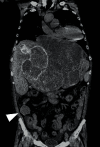
(a) Plain abdominal CT showing an irregular high-density area in the center of the liver and bleeding is suspected. Tumor detection is difficult. (b) Contrast-enhanced CT (arterial phase) revealing multiple tumors with a contrast effect in the left lobe of the liver (arrow). The patient was diagnosed with tumor-associated hemorrhage. (c) Abdominal CT (late phase) disclosing that the tumor exhibits the same contrast effect as normal liver tissue.
Figure 2
Figure 2
(a) Diffusion-weighted MRI images showing multiple tumors with diffusion decrease in the left lobe of the liver. (b) On MRCP, the intrahepatic bile duct was observed to be markedly compressed by bleeding in the hilum of the liver.
Figure 3
Figure 3
Abdominal ultrasonography showing a 15-mm hypoechoic mass in liver segment S2 adjacent to the bleeding (arrowhead). A percutaneous biopsy was performed.
Figure 4
Figure 4
Pathological analysis of the liver biopsy revealed dense proliferation of spindle-shaped atypical cells. The tumor cells were positive for c-kit and CD34, and the patient was diagnosed with GIST. The MIB-1 labelling index was 2% (x 400).
Figure 5
Figure 5
CT findings one month before death. Multiple metastases of GIST are found in the liver. A 40 mm nodule is found in the ileocecal region (arrowhead). No tumor was found in both lungs.
Figure 6
Figure 6
On autopsy, a tumor with necrotic hemorrhage extending throughout the liver could be seen macroscopically, with only little normal liver tissues remaining.
Figure 7
Figure 7
Pathological findings in the liver tumor at autopsy. Proliferation of atypical spindle-shaped cells was observed. The tumor was c-kit- and CD34-positive, confirming the diagnosis of GIST. MIB-1 labelling index was 12% (x 400).
Figure 8
Figure 8
On autopsy, a 40-mm nodule was found in the ileocecal region, and the pathological finding was compatible with GIST. The patient was diagnosed with peritoneal dissemination of GIST.
Figure 9
Figure 9
Lung findings at autopsy. (a) Macroscopic findings did not indicate a clear tumor in the lungs (x100). (b, c) Microscopically, multiple small lung metastases of GIST were observed in all lung lobes (arrow) (x200, Hematoxylin-Eosin stain).

References

    1. DeMatteo R. P., Lewis J. J., Leung D., Mudan S. S., Woodruff J. M., Brennan M. F. Two hundred gastrointestinal stromal tumors. Annals of Surgery. 2000;231(1):51–58. doi: 10.1097/00000658-200001000-00008. - DOI - PMC - PubMed
    1. Koo D.-H., Ryu M.-H., Kim K.-M., et al. Asian consensus guidelines for the diagnosis and management of gastrointestinal stromal tumor. Cancer Research and Treatment. 2016;48(4):1155–1166. doi: 10.4143/crt.2016.187. - DOI - PMC - PubMed
    1. Hirota S., Isozaki K., Moriyama Y., et al. Gain-of-function mutations of c-kit in human gastrointestinal stromal tumors. Science. 1998;279(5350):577–580. doi: 10.1126/science.279.5350.577. - DOI - PubMed
    1. Roberts P. J., Eisenberg B. Clinical presentation of gastrointestinal stromal tumors and treatment of operable disease. European Journal of Cancer. 2002;38:S37–S38. doi: 10.1016/s0959-8049(02)80601-3. - DOI - PubMed
    1. Sturgeon C., Chejfec G., Espat N. J. Gastrointestinal stromal tumors: a spectrum of disease. Surgical Oncology. 2003;12(1):21–26. doi: 10.1016/s0960-7404(02)00074-9. - DOI - PubMed

Publication types

LinkOut - more resources