. 2021 Jan 20;12(2):955-968.
doi: 10.1364/BOE.402081. eCollection 2021 Feb 1.

Colon phantoms with cancer lesions for endoscopic characterization with optical coherence tomography

Affiliations

Colon phantoms with cancer lesions for endoscopic characterization with optical coherence tomography

Natalia Zulina et al. Biomed Opt Express. .

Abstract

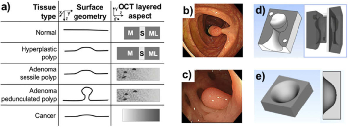
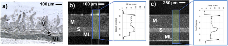
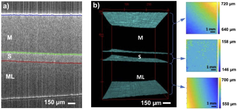
Optical coherence tomography (OCT) is a growing imaging technique for real-time early diagnosis of digestive system diseases. As with other well-established medical imaging modalities, OCT requires validated imaging performance and standardized test methods for performance assessment. A major limitation in the development and testing of new imaging technologies is the lack of models for simultaneous clinical procedure emulation and characterization of healthy and diseased tissues. Currently, the former can be tested in large animal models and the latter can be tested in small animal disease models or excised human biopsy samples. In this study, a 23 cm by 23 cm optical phantom was developed to mimic the thickness and near-infrared optical properties of each anatomical layer of a human colon, as well as the surface topography of colorectal polyps and visual appearance compatible with white light endoscopy.


Conflict of interest statement

The authors declare no conflicts of interest.

Figures

Fig. 1.
Fig. 1.
(a) Schematic representation of design inputs for surface geometry and inner tissue architecture for most frequent tissue types [42]; Endoscopic image of (b) pedunculated polyp and (c) sessile polyp adapted with permission from [44]; CAD drawings of a mold for pedunculated polyp (d) and sessile polyp (e) phantom.
Fig. 2.
Fig. 2.
Process diagram for the fabrication of a colon phantom with flat cancerous lesion, pedunculated and sessile polyps.
Fig. 3.
Fig. 3.
(a) Unfolded 23 cm by 23 cm colon phantom with cancerous insertions and benign polyps (before blood vessels drawing). 3D-printed VeroWhite molds for (b) sessile and (c) pedunculated polyp phantoms. (d) internal and (e) external views of the folded colon phantom.
Fig. 4.
Fig. 4.
(a) Histology of a swine bowel tissue. OCT images and intensity plots of 50 averaged A-scans of (b) swine bowel ex vivo and (c) phantom. Scale bar represents a physical size.
Fig. 5.
Fig. 5.
Volumetric rendering of 3D OCT data and cross-sectional OCT images of different tissue types present in the phantom obtained with a custom benchtop imaging system and compared with OCT images of corresponding tissue types obtained in human adapted with permission from [50,54]. The volumetric data for the pedunculated polyp was acquired from its side to better present its shape.
Fig. 6.
Fig. 6.
Thickness analysis of a 6 mm by 6 mm area of the phantom mimicking normal tissue. (a) Segmentation of 2D OCT image with mucosa (M) in between blue and green lines, submucosa (S) between green and red lines and muscularis layer (ML) between red and black line. (b) Rreconstructed 3D view of automatically segmented layers and corresponding 2D thickness maps of mucosa (top), submucosa (middle) and muscularis layer (bottom).
Fig. 7.
Fig. 7.
White light endoscopy and OCT examination of the colon model. (a) External view of the assembled colon phantom. (b) Picture of the OCT catheter inserted in the working channel of the endoscope. (c) Internal view of phantom with visible lesions and folds during retroflexion examination inside the phantom obtained with a 150° turn of the tip of the endoscope. Endoscopic images and corresponding OCT cross-sections of (d) healthy tissue with layered architecture of mucosa (M), submucosa (S) and muscular layer (ML), (e) non neoplastic mucosal growth phantom, representing a benign lesion with visible thickening of the mucosa (yellow arrows), (f) pedunculated polyp phantom, and (g) flat cancerous tissue (CT) on the left, next to the healthy tissue (HT) sessile.
Fig. 8.
Fig. 8.
Image quality analysis showing: (a) background intensity distribution, (b) SNR and (c) local contrast of the OCT phantom images obtained with the custom catheter and a bench-top mode, compared to OCT images of the human colon from a commercial volumetric laser endomicroscopy (VLE).

Similar articles

Cited by

References

    1. “Cancer,” https://www.who.int/news-room/fact-sheets/detail/cancer.
    1. Tsai T.-H., Fujimoto J. G., Mashimo H., “Endoscopic Optical Coherence Tomography for Clinical Gastroenterology,” Diagnostics 4(2), 57–93 (2014).10.3390/diagnostics4020057 - DOI - PMC - PubMed
    1. Gora M. J., Sauk J. S., Carruth R. W., Gallagher K. A., Suter M. J., Nishioka N. S., Kava L. E., Rosenberg M., Bouma B. E., Tearney G. J., “Tethered capsule endomicroscopy enables less invasive imaging of gastrointestinal tract microstructure,” Nat. Med. 19(2), 238–240 (2013).10.1038/nm.3052 - DOI - PMC - PubMed
    1. Smith G. T., Lurie K. L., Zlatev D. V., Liao J. C., Ellerbee Bowden A. K., “Multimodal 3D cancer-mimicking optical phantom,” Biomed. Opt. Express 7(2), 648–662 (2016).10.1364/BOE.7.000648 - DOI - PMC - PubMed
    1. Jäckle S., Gladkova N., Feldchtein F., Terentieva A., Brand B., Gelikonov G., Gelikonov V., Sergeev A., Fritscher-Ravens A., Freund J., Seitz U., Soehendra S., Schrödern N., “In vivo endoscopic optical coherence tomography of the human gastrointestinal tract–toward optical biopsy,” Endoscopy 32(10), 743–749 (2000).10.1055/s-2000-7711 - DOI - PubMed

LinkOut - more resources