Intraductal Papillary Neoplasm of the Bile Duct: A Rare Case of Intrahepatic Space-Occupying Lesion
- PMID: 33680605
- PMCID: PMC7929547
- DOI: 10.7759/cureus.13063
Intraductal Papillary Neoplasm of the Bile Duct: A Rare Case of Intrahepatic Space-Occupying Lesion
Abstract
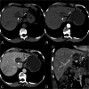
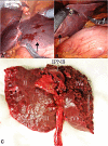
Intraductal papillary neoplasm of the bile duct (IPNB) is a rare tumor and is considered one of the precursor lesions for cholangiocarcinoma. Though relatively common in the far east countries, it is uncommon in the Indian population. A 67-year-old gentleman presented with vague upper abdominal pain with no history of fever, jaundice, melena, or hematemesis. An abdominal ultrasound showed a solid cystic lesion in the left lobe of the liver with upstream dilatation of bile ducts. Computed tomography and magnetic resonance imaging showed similar findings. With a differential diagnosis of intrahepatic cholangiocarcinoma, intraductal papillary neoplasm, and biliary cystadenoma, he underwent robotic-assisted left hepatectomy. Histopathology was suggestive of IPNB. Following surgery, he had an uneventful recovery and was advised for follow-up visits every six months. A clinical, radiological, and pathological profile of this rare tumor has been described here with a review of the existing literature.
Keywords: biliary papillomatosis; bt-ipmn; cholangiocarcinoma; intraductal papillary neoplasm of bile duct; ipnb.
Copyright © 2021, Dutta et al.
Conflict of interest statement
The authors have declared that no competing interests exist.
Figures


Similar articles
-
Intraductal Papillary Neoplasm of the Bile Duct: Radiological Diagnosis of a Rare Entity: Case Series.Euroasian J Hepatogastroenterol. 2023 Jan-Jun;13(1):28-31. doi: 10.5005/jp-journals-10018-1385. Euroasian J Hepatogastroenterol. 2023. PMID: 37554972 Free PMC article.
-
Intraductal papillary neoplasm of the bile duct presenting with hepatogastric fistula: a case report and literature review.Front Oncol. 2023 May 19;13:1193918. doi: 10.3389/fonc.2023.1193918. eCollection 2023. Front Oncol. 2023. PMID: 37274235 Free PMC article.
-
Branch-type intraductal papillary neoplasm of the bile duct treated with laparoscopic anatomical resection: a case report.Surg Case Rep. 2020 May 15;6(1):103. doi: 10.1186/s40792-020-00864-3. Surg Case Rep. 2020. PMID: 32415464 Free PMC article.
-
Intraductal papillary neoplasm of the bile duct: review of updated clinicopathological and imaging characteristics.Br J Surg. 2023 Aug 11;110(9):1229-1240. doi: 10.1093/bjs/znad202. Br J Surg. 2023. PMID: 37463281 Review.
-
Intraductal papillary neoplasm of the bile ducts: A case report and literature review.World J Gastroenterol. 2015 Nov 21;21(43):12498-504. doi: 10.3748/wjg.v21.i43.12498. World J Gastroenterol. 2015. PMID: 26604656 Free PMC article. Review.
Cited by
-
Surgical Treatment of Intraductal Papillary Neoplasm of the Bile Duct: A Report of Two Cases and Review of the Literature.Front Oncol. 2022 Jun 23;12:916457. doi: 10.3389/fonc.2022.916457. eCollection 2022. Front Oncol. 2022. PMID: 35814451 Free PMC article.
References
-
- Precancerous lesions of the biliary tree. Klöppel G, Adsay V, Konukiewitz B, Kleeff J, Schlitter AM, Esposito I. Best Pract Res Clin Gastroenterol. 2013;27:285–297. - PubMed
-
- Mucinous cystic neoplasm of the liver or intraductal papillary mucinous neoplasm of the bile duct? A case report and a review of literature. Kunovsky L, Kala Z, Svaton R, Moravcik P, Mazanec J, Husty J, Prochazka V. Ann Hepatol. 2018;17:519–524. - PubMed
-
- Intraductal papillary neoplasm of the bile duct: a rarity. Bal M, Goel M, Ramadwar M, Deodhar K. Indian J Pathol Microbiol. 2014;57:144–145. - PubMed
-
- Biliary tract intraductal papillary mucinous neoplasm: a brief report and review of literature. Subhash R, Valiyaveettil IA, Natesh B, Raji L. Indian J Pathol Microbiol. 2014;57:588–590. - PubMed
Publication types
LinkOut - more resources
Full Text Sources
Other Literature Sources
