. 2021 Feb 22:2021:6634429.
doi: 10.1155/2021/6634429. eCollection 2021.

Ferrotoxicity and Its Amelioration by Calcitriol in Cultured Renal Cells

Affiliations

Ferrotoxicity and Its Amelioration by Calcitriol in Cultured Renal Cells

Chandrashekar Annamalai et al. Anal Cell Pathol (Amst). .

Abstract

Globally, acute kidney injury (AKI) is associated with significant mortality and an enormous economic burden. Whereas iron is essential for metabolically active renal cells, it has the potential to cause renal cytotoxicity by promoting Fenton chemistry-based oxidative stress involving lipid peroxidation. In addition, 1,25-dihydroxyvitamin D3 (calcitriol), the active form of vitamin D, is reported to have an antioxidative role. In this study, we intended to demonstrate the impact of vitamin D on iron-mediated oxidant stress and cytotoxicity of Vero cells exposed to iohexol, a low osmolar iodine-containing contrast media in vitro. Cultured Vero cells were pretreated with 1,25-dihydroxyvitamin D3 dissolved in absolute ethanol (0.05%, 2.0 mM) at a dose of 1 mM for 6 hours. Subsequently, iohexol was added at a concentration of 100 mg iodine per mL and incubated for 3 hours. Total cellular iron content was analysed by a flame atomic absorption spectrophotometer at 372 nm. Lipid peroxidation was determined by TBARS (thiobarbituric acid reactive species) assay. Antioxidants including total thiol content were assessed by Ellman's method, catalase by colorimetric method, and superoxide dismutase (SOD) by nitroblue tetrazolium assay. The cells were stained with DAPI (4',6-diamidino-2-phenylindole), and the cytotoxicity was evaluated by viability assay (MTT assay). The results indicated that iohexol exposure caused a significant increase of the total iron content in Vero cells. A concomitant increase of lipid peroxidation and decrease of total thiol protein levels, catalase, and superoxide dismutase activity were observed along with decreased cell viability in comparison with the controls. Furthermore, these changes were significantly reversed when the cells were pretreated with vitamin D prior to incubation with iohexol. Our findings of this in vitro model of iohexol-induced renotoxicity lend further support to the nephrotoxic potential of iron and underpin the possible clinical utility of vitamin D for the treatment and prevention of AKI.


Conflict of interest statement

The authors declare that they have no conflicts of interest.

Figures

Figure 1
Figure 1
MTT 3-(4,5-dimethyl (thiazol-2-yl)-2,5-diphenyl tetrazolium bromide) assay to quantify cell viability in Vero cells exposed to graded concentrations of iohexol. One-way ANOVA and Tukey posttest (n = 4) were used to analyse the results. p < 0.05, ∗∗p < 0.01, and ∗∗∗p < 0.001 (2-tailed) in comparison with the control.
Figure 2
Figure 2
Vero cells under the microscope after subjecting to different doses of iohexol containing increasing concentration of iodine (I) (n = 4). Magnification 100x. The control group displays healthy, growing cells with elongated morphology (black arrows), whereas the cells incubated with iohexol show signs of rounding up.
Figure 3
Figure 3
Relative concentrations of total cellular iron determined by atomic absorption spectroscopy in Vero cells treated with iohexol at a concentration of 100 mg iodine/mL or pretreated with 1 μM vitamin D3. Results are represented as mean ± SD. One-way ANOVA and Tukey posttest (n = 4) were used to analyse the results. ns: not significant; p < 0.05, ∗∗p < 0.01, and ∗∗∗p < 0.001 (2-tailed) in comparison with the control; $p < 0.05, $$p < 0.01, and $$$p < 0.001 (2-tailed) iohexol (I) versus iohexol+vitamin D (I+Vit D) groups.
Figure 4
Figure 4
Levels of (a) lipid peroxidation, (b) thiol protein, (c) catalase, and (d) superoxide dismutase activity in Vero cells treated with iohexol at a concentration of 100 mg iodine/mL or pretreated with 1 μM vitamin D3. Results are represented as mean ± SD. One-way ANOVA and Tukey posttest (n = 4) were used to analyse the results. ns: not significant; p < 0.05, ∗∗p < 0.01, and ∗∗∗p < 0.001 (2-tailed) versus control; $p < 0.05, $$p < 0.01, and $$$p < 0.001 (2-tailed) iohexol (I) versus iohexol+vitamin D (I+Vit D) groups.
Figure 5
Figure 5
Fluorescent micrographs of Vero cells stained with DAPI (4′,6-diamidino-2-phenylindole) demonstrating more prominent nuclear condensation (shown in blue) in the iohexol-treated (I100) group (white arrows) compared to the iohexol+vitamin D-treated (Vit D+I100) group. Magnification 100x.
Figure 6
Figure 6
MTT 3-(4,5-dimethyl (thiazol-2-yl)-2,5-diphenyl tetrazolium bromide) assay to quantify cell viability in Vero cells exposed to iohexol and vitamin D3. Results are represented as mean ± SD. One-way ANOVA and Tukey posttest (n = 4) were used to analyse the results. ns: not significant; p < 0.05, ∗∗p < 0.01, and ∗∗∗p < 0.001 (2-tailed) in comparison with the control; $p < 0.05, $$p < 0.01, and $$$p < 0.001 (2-tailed) iohexol (I) versus iohexol+vitamin D (I+Vit D) groups.
Figure 7
Figure 7
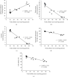
Scatter plot depicting the correlation between total cellular iron, oxidative stress, and cell viability in cells treated with iohexol: (a) lipid peroxidation, (b) thiol protein, (c) catalase, (d) superoxide dismutase activity, and (e) cell viability. NBT: nitroblue tetrazolium.
Figure 8
Figure 8
Mechanistic diagram illustrating the renoprotective role of calcitriol in iohexol-induced AKI. Iohexol increases the intracellular release of catalytic iron, for example, from mitochondria (MT) during the process of renal tissue injury. Concurrently, 1 α-hydroxylase (CYPB21) catalyses the conversion of 25-hydroxyvitamin D3 (25[OH]D) to active 1,25-dihydroxyvitamin D3 (I,25[OH]2D3), also known as calcitriol. Calcitriol binds to vitamin D receptor (VDR) which then binds to the proximal promoter region of HAMP gene containing vitamin D-responsive elements (VDREs) leading to suppression of HAMP gene and therefore hepcidin protein expression directly [112]. Vitamin D also indirectly reduces prohepcidin inflammatory cytokines, IL-6 and IL-1β [113] and MCP-1 [114]. Furthermore, vitamin D possesses antioxidant property and it relieves endoplasmic reticulum stress which is an inducer of hepcidin expression that could result in dysregulated cellular iron homeostasis [115]. Transferrin-bound iron (TBI) enters the cytosol through transferrin receptor protein (TfR1 and TfR2) and is either oxidized by ferritin heavy chain (FtH) to be stored intracellularly in the ferritin complex consisting of ferritin heavy and light chains (FtH/FtL) or is exported out of the cell by ferroportin (FPN). Hepcidin, the key regulator of systemic and cellular iron homeostasis, facilitates internalization and degradation of ferroportin. 25[OH]D: 25-hydoxyvitamin D3; 1,25[OH]2D: 1,25-dihyroxyvitamin D3; VDR: vitamin D receptor; MCP-1: monocyte chemoattractant protein 1; IL-6: interleukin 6; IL-1β: interleukin 1β; IL-6R: interleukin 6 receptor; IL-1βR: interleukin 1β receptor; STAT RE: signal transducer and activator of transcription responsive element; HAMP: hepcidin antimicrobial peptide; ER stress: endoplasmic reticulum stress; MT: mitochondria; Fe(III): ferric iron; Tf: transferrin; TBI: transferrin-bound iron; TfR1: transferrin receptor protein 1; TfR2: transferrin receptor protein 2; Fe(II): ferrous iron; FtH/FtL: ferritin heavy chain, ferritin light chain; FPN: ferroportin.

Similar articles

Cited by

References

    1. Li P. K. T., Burdmann E. A., Mehta R. L., Martin S. Acute kidney injury: global health alert. Journal of Nephropathology. 2013;2(2):90–97. doi: 10.5812/nephropathol.10449. - DOI - PMC - PubMed
    1. Lakhmir S., Chawla, on behalf of the Acute Disease Quality Initiative Workgroup 16, Bellomo R., et al. Acute kidney disease and renal recovery: consensus report of the acute disease quality initiative (ADQI) 16 workgroup. Nature Reviews Nephrology. 2017;13(4):241–257. doi: 10.1038/nrneph.2017.2. - DOI - PubMed
    1. Kinsey G. R., Okusa M. D. Pathogenesis of acute kidney injury: foundation for clinical practice. American Journal of Kidney Diseases: The Official Journal of the National Kidney Foundation. 2011;58(2):291–301. doi: 10.1053/j.ajkd.2011.02.385. - DOI - PMC - PubMed
    1. Bonventre J. V. Pathophysiology of AKI: injury and normal and abnormal repair. Contributions to Nephrology. 2010;165:9–17. doi: 10.1159/000313738. - DOI - PubMed
    1. Bagshaw S. M., Wald R. Acute kidney injury: timing of renal replacement therapy in AKI. Nature Reviews Nephrology. 2016;12(8):445–446. doi: 10.1038/nrneph.2016.92. - DOI - PubMed

LinkOut - more resources