. 2021 Jan 30;10(2):e437-e450.
doi: 10.1016/j.eats.2020.10.028. eCollection 2021 Feb.

Latarjet Cerclage: The All-Arthroscopic Metal-Free Fixation

Affiliations

Latarjet Cerclage: The All-Arthroscopic Metal-Free Fixation

Abdul-Ilah Hachem et al. Arthrosc Tech. .

Abstract

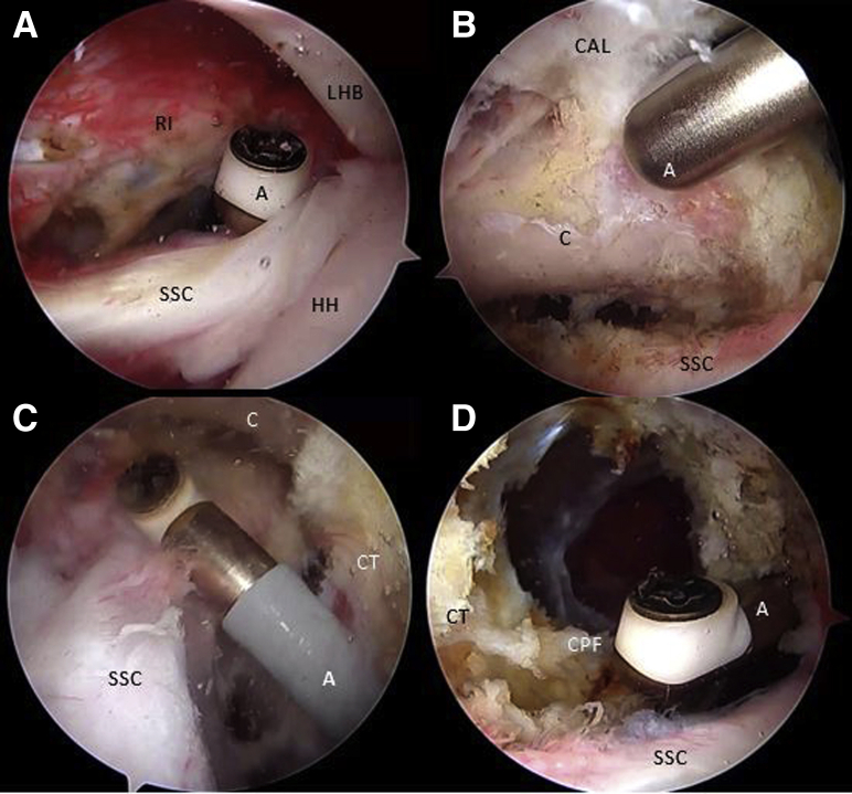
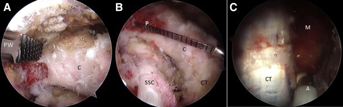
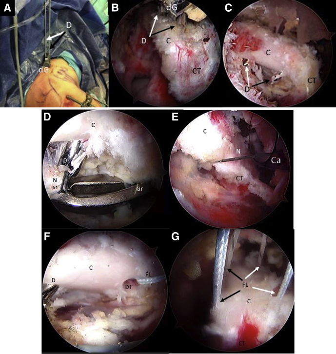
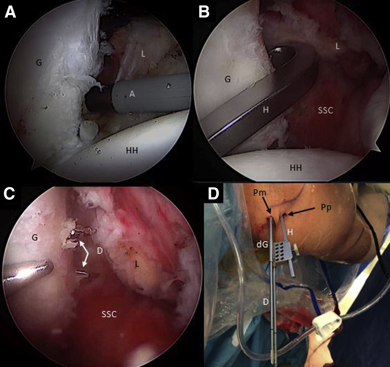
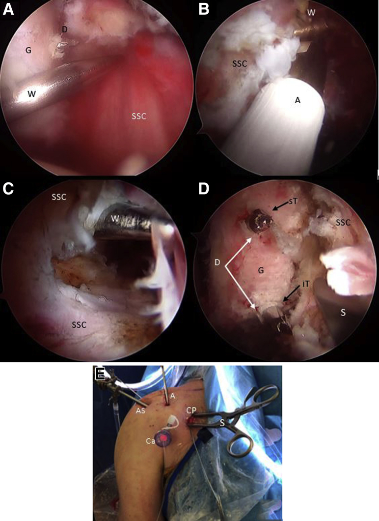
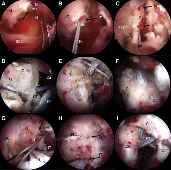
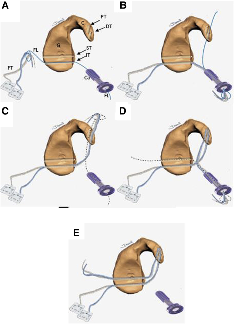
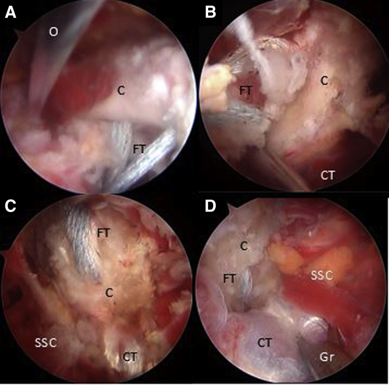
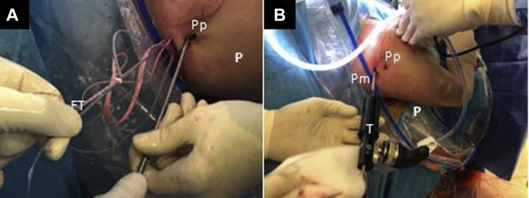
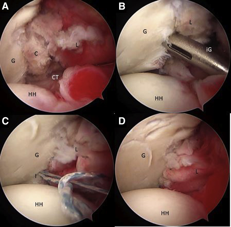
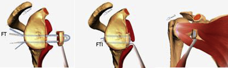
The Bristow-Latarjet procedure has been one of the most recognized procedures for the treatment of recurrent shoulder dislocation with anterior glenoid bone loss, revision surgery after failed Bankart repair, contact and collision sport injuries, and patients with a high risk of recurrence. Open and arthroscopic approaches have recently shown similar outcomes by several authors. However, complications related to metal implants, despite being low, are still a matter of concern. We describe an all-arthroscopic Latarjet technique with a metal-free fixation method using 2 ultra-high-strength sutures, creating a cerclage construct through 2.4mm glenoid and coracoid tunnels with a final capsulolabral complex reconstruction.


Figures

Fig 1
Fig 1
External view of the (A) preoperative setting with the patient placed in the beach-chair position with the right arm fixed on a Trimano Arthrex device and (B) drawing of bony structures and arthroscopic portals. (A, anterior portal; AS, anterosuperior portal; AI, anteroinferior portal; CP, coracoid portal.)
Fig 2
Fig 2
Arthroscopic view of a right shoulder through posterior portal with the patient placed in beach-chair position, showing an engaging lesion between the HS lesion and the anterior glenoid bone defect with the arm in abduction and external rotation. (G, glenoid surface; HH, humeral head; HS, Hill–Sachs lesion.)
Fig 3
Fig 3
Right shoulder, Beach-chair position, Arthroscopic view, 30° scope. (A) The rotator interval is released looking through the posterior portal and working through the anterosuperior portal with an ablator. (B) The coracoacromial ligament is released through the anterosuperior portal with an ablator and looking through the posterior portal. (C) The superior and presubscapularis tendon space is released with an ablator looking through the anterosuperior portal and working through the anteroinferior portal. (D) The clavipectoral fascia is released with an ablator looking through the anterosuperior portal and working through the anteroinferior portal. (A, ablator; C, coracoid; CAL, coracoacromial ligament; CPF, clavipectoral fascia; CT, conjoint tendon; HH, humeral head; LHB, long head of the biceps tendon; RI, rotator interval; SSC, subscapularis tendon.)
Fig 4
Fig 4
Right shoulder, beach-chair position, Arthroscopic view, 30° scope. (A) The coracoid is shaped on its lateral and inferior aspect looking through the anterior portal and working through the anterosuperior portal with a PowerRasp (Arthrex). (B) The length of the coracoid is measured with an Arthroscopic Probe (Arthrex) from the anterior portal and looking through the anterosuperior portal. (C) Looking through the anterosuperior portal, the pectoralis minor tendon is released from the medial aspect of the coracoid. (A, ablator; C, coracoid; CT, conjoint tendon; M, medial coracoid space; P, arthroscopic measurement probe; PW, PowerRasp; SSC, subscapularis tendon.)
Fig 5
Fig 5
Right shoulder, beach-chair position. (A) Beach-chair position, right shoulder, Anterosuperior extraarticular view of the drilling guide inserted through the coracoid portal. (B) Arthroscopic view, 70° scope, looking through the anterosuperior portal, drilling guide inserted through the coracoid portal and (C) 2 cannulated drills, can be observed. Note: In (A-C), black arrows show the distal coracoid tunnel and white arrows show the proximal coracoid tunnel. (D-F) Arthroscopic view, 70° scope, looking through the anterosuperior portal, a nitinol wire is retrieved from the distal coracoid tunnel through the cannula in the anteroinferior portal and then replaced with a with a blue FiberLink. (G) The procedure is repeated with the proximal coracoid tunnel, exchanging the nitinol wire with a different-colored FiberLink than in the distal tunnel. Then all FiberLink tails are retrieved through the coracoid portal. Note: white arrows show FiberLink strands that are passing through coracoid tunnels. Black arrows show FiberLink strands that are passing medial to the coracoid process. (C, coracoid process; Ca, cannula; CT, conjoint tendon; D, cannulated drill; dG, drilling guide; DT, distal coracoid tunnel; FL, FiberLink; Gr, grasper; N, nitinol wire.)
Fig 6
Fig 6
Right shoulder, beach chair position, anterosuperior portal, arthroscopic view, 70° scope. (A) The anterior capsulolabral complex is released with an ablator through the coracoid portal. (B) The hook component of the drilling guide is inserted through the posterior portal and engaged in the anteroinferior glenoid neck. (C) Then, the hook is connected with the drilling guide outside the patient and pushed against the posterior glenoid neck through a separate posteromedial portal. Two cannulated drills are inserted through the glenoid drilling guide in the posteromedial portal to perform the glenoid tunnels. (D) Posterosuperior external view of the right shoulder in the beach chair position showing the drilling guide construct insertion through the posterior and the posteromedial portals. (A, ablator; D, cannulated drill; dG, drilling guide; G, glenoid; H, hook component of the drilling construct; HH, humeral head; L, capsulolabral complex; Pm, posteromedial portal; Pp, posterior portal; SSC, subscapularis muscle.)
Fig 7
Fig 7
Right shoulder, beach-chair position, arthroscopic view, 70° scope, anterosuperior portal. (A) The Wissinger is inserted through the posterior portal to determine the split level of the subscapularis tendon. (B-C) The Wissinger rod is identified across the anterior aspect of the subscapularis muscle and the split is performed parallel to the muscle fibers with an ablator. (D) Deep to the subscapularis split, the glenoid and the cannulated drills are identified. A specific spreader is inserted through the coracoid portal to facilitate anterior glenoid neck visualization and sutures shuttling. (E) Anterior external view of the right shoulder in the beach-chair position showing the spreader insertion through the coracoid portal and the cannula insertion in the anteroinferior portal for subsequent sutures shuttling. (A, ablator; AP, anterior portal; AS, anterosuperior portal; C, coracoid process; Ca, cannula; CP, coracoid portal; D, cannulated drill; G, glenoid; iT, inferior glenoid tunnel; S, self-retaining retractor; SSC, subscapularis muscle; sT, superior glenoid tunnel; W, Wissinger rod.)
Fig 8
Fig 8
Right shoulder, beach-chair position, arthroscopic view, 70° scope, anterosuperior portal. (A-C) With an arthroscopic grasper, a nitinol wire is retrieved from the inferior glenoid tunnel, through the subscapularis muscle split and the cannula in the anteroinferior portal. The nitinol is replaced with a FiberLink and then subsequently with the FiberTape from the posterior to the anterior side of the glenoid. (D-F) By pulling the blue distal coracoid FiberLink assembled with the FiberTape through the cannula outside the patient, these are shuttled from the inferior to the superior side of the coracoid (distal coracoid drill hole). In the same way, the white FiberLink is pulled through the cannula to shuttle the FiberTape from the superior to the inferior side of the coracoid process (proximal drill hole). (G-I) With an arthroscopic grasper, a nitinol wire is retrieved from the superior glenoid tunnel, through the subscapularis muscle split and the cannula in the anteroinferior portal. The nitinol is replaced with a FiberLink. Then the FiberLink, previously connected with the FiberTape outside the patient, is pulled from the cannula from the anterior side to the posterior side of the glenoid. (C, coracoid process; Ca, cannula; CT, conjoint tendon; D, cannulated drill; FL, FiberLink; FT, FiberTape; G, glenoid; Gr, arthroscopic grasper; iT, inferior glenoid tunnel; N, nitinol wire; SSC, subscapularis muscle; sT, superior glenoid tunnel.)
Fig 9
Fig 9
Schematic representation of sutures shuttling technique through the glenoid and coracoid process sequentially from figure A to E. The inbound-outbound shuttling process is divided in 4 phases. The FiberTape must be equally tensioned at the same length in every shuttling phase. Inbound process: (A) Phase 1: From the posterior to the anterior side of the glenoid, the FiberTapes are transferred through the inferior glenoid tunnel, across the subscapularis muscle split and retrieved through the cannula at the anteroinferior portal. (B) Phase 2: The FiberTapes are transferer from the cannula through the distal coracoid tunnel from ventral to dorsal. Outbound process: (C) Phase 3: The FiberTapes are shuttled from the dorsal to the ventral side of the coracoid and retrieved through the cannula. (D) Phase 4: The FiberTapes are shuttled from the cannula through the subscapularis split and pulled from the anterior side to the posterior side of the glenoid. (E) Schematic representation of the final construct before performing the coracoid osteotomy. (C, coracoid; Ca, cannula; DT, distal coracoid tunnel; FT, FiberTape; FL, FiberLink; G, glenoid; IT, inferior glenoid tunnel; PT, proximal coracoid tunnel; ST, superior glenoid tunnel.)
Fig 10
Fig 10
Right shoulder, beach-chair position, arthroscopic view, 70° scope, anterosuperior portal. An arthroscopic burr and chisel are used to osteotomized the coracoid, inserted through the anterior portal. (B-D) By Pulling the FiberTapes, the coracoid is transported through the subscapularis muscle split to the anterior glenoid neck. An arthroscopic grasper is used to facilitate the coracoid passage across the subscapularis split. (C, coracoid; CT, conjoint tendon; FT, FiberTape; Gr, grasper; O, chisel; SSC, subscapularis muscle.)
Fig 11
Fig 11
Posterior extraarticular view of the right shoulder, beach-chair position. (A) The FiberTape Cerclage System is interconnected, and traction is performed subsequently one by one for each strand to fix the coracoid in the anterior glenoid neck. (B) A tensioner (Arthrex) is used to achieve a strong fixation of up to 100 N. (FT: FiberTape; P, posterior right shoulder; Pm, posteromedial portal; Pp, posterior portal; T, tensioner.).
Fig 12
Fig 12
(A-D) Right shoulder, beach-chair position, arthroscopic view, 70° scope, anterosuperior portal. The capsulolabral complex is repaired and fixed in the native anterior glenoid rim with 3 knotless FiberTak 1.8 soft anchors (Arthrex) from the anterior portal. (C, coracoid; G, glenoid; L, capsulolabral complex; HH, humeral head; I, implant; iG, implant guide.)
Fig 13
Fig 13
Schematic representation of the final Latarjet cerclage construct. The circle-like configuration of the fixation can be noted. FiberTape cerclage sutures (FT), FiberTape interconnected (FTI).

References

    1. Latarjet M. Treatment of recurrent dislocation of the shoulder. Lyon Chir. 1954;49:994–997. - PubMed
    1. Hurley E.T., Jamal M.S., Ali Z.S., Montgomery C., Pauzenberger L., Mullett H. Long-term outcomes of the Latarjet procedure for anterior shoulder instability: A systematic review of studies at 10-year follow-up. J Shoulder Elbow Surg. 2019;28:e33–e39. - PubMed
    1. Young A.A., Maia R., Berhouet J., Walch G. Open Latarjet procedure for management of bone loss in anterior instability of the glenohumeral joint. J Shoulder Elbow Surg. 2011;20:S61–S69. - PubMed
    1. Provencher M.T., Bhatia S., Ghodadra N.S. Recurrent shoulder instability: Current concepts for evaluation and management of glenoid bone loss. J Bone Joint Surg A. 2010;92(suppl 2):133–151. - PubMed
    1. Provencher M.T., Ferrari M.B., Sanchez G., Anavian J., Akamefula R., Lebus G.F. Current treatment options for glenohumeral instability and bone loss. JBJS Rev. 2017;5:1–11. - PubMed