

Evaluation of the degenerative pattern of PCL in osteoarthritis patients using UTE-T2 mapping
- PMID: 33680861
- PMCID: PMC7899951
- DOI: 10.1016/j.asmart.2021.01.004
Evaluation of the degenerative pattern of PCL in osteoarthritis patients using UTE-T2 mapping
Abstract
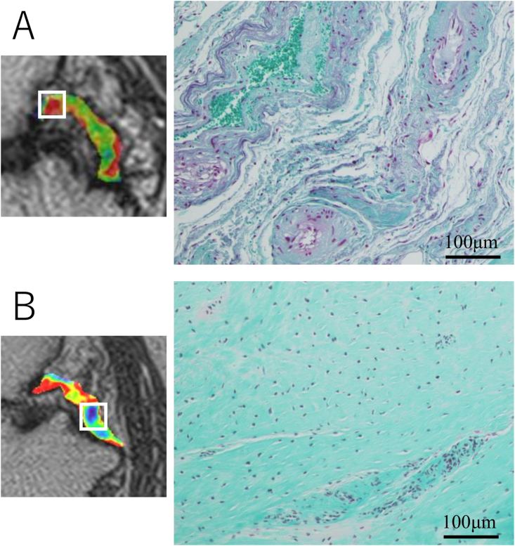
Background: The posterior cruciate ligament (PCL) is one of the essential stabilizers of the knee joint and it was demonstrated that its degenerative change related to the knee osteoarthritis (OA). We aimed to evaluate signal of the PCL in OA patients in comparison with healthy young and elderly volunteers using the ultra-short echo timeenhanced (UTE)-T2∗ mapping, and to validate these findings with histology.
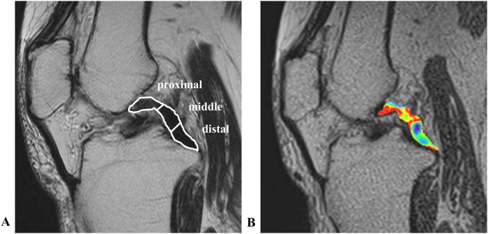
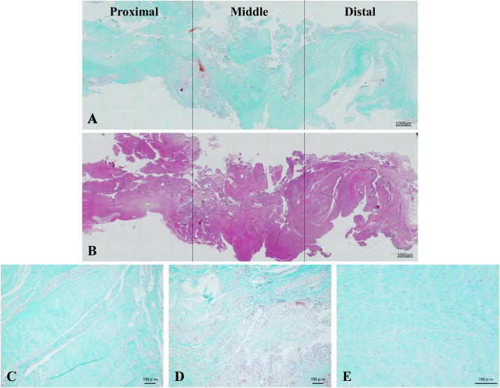
Methods: Thirty asymptomatic volunteers, 13 young people (younger group) and 17 elderly people (elder group), and 27 patients who had undergone total knee arthroplasty (OA group) were enrolled in this study. UTE-T2∗ maps of PCL were obtained from all participants. The PCL was divided into proximal, middle, and distal parts and the UTET2∗ values obtained from each part were compared among the groups. In OA group, the sacrificed PCLs were evaluated histologically in each part corresponding to the part of UTE-T2∗ maps and compared.
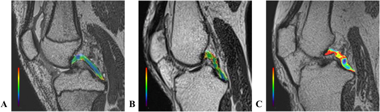
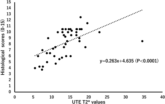
Results: The UTE-T2∗ values in OA group were significantly higher than those in other groups except in distal part. In elder group, the UTE-T2∗ values were significantly higher than those in younger group only in the proximal part. Moreover, in OA group, the UTE-T2∗ values in proximal and middle parts were significantly higher than those in distal part. There was a moderate correlation between the UTE-T2∗ values and histological scores.
Conclusions: The specific signal intensity pattern of the PCL in patients with OA was demonstrated using UTE-T2∗ mapping, and these findings were related to histological degenerated status of the PCL.
Keywords: Magnetic resonance image (MRI); Osteoarthritis (OA); Posterior cruciate ligament (PCL); Ultra-short echo time-enhanced (UTE).
© 2021 Asia Pacific Knee, Arthroscopy and Sports Medicine Society. Published by Elsevier (Singapore) Pte Ltd.?.
Figures
References
-
- Creamer P., Hochberg M.C. Osteoarthritis. Lancet. 1997;350(9076):503–508. - PubMed
-
- Dieppe P.A., Lohmander L.S. Pathogenesis and management of pain in osteoarthritis. Lancet. 2005;365(9463):965–973. - PubMed
-
- LaPrade C.M., Civitarese D.M., Rasmussen M.T., LaPrade R.F. Emerging updates on the posterior cruciate ligament: a review of the current literature. Am J Sports Med. 2015;43(12):3077–3092. - PubMed
LinkOut - more resources
Full Text Sources
Other Literature Sources
