Biocompatibility and bioactive potential of the NeoMTA Plus endodontic bioceramic-based sealer
- PMID: 33680893
- PMCID: PMC7906839
- DOI: 10.5395/rde.2021.46.e4
Biocompatibility and bioactive potential of the NeoMTA Plus endodontic bioceramic-based sealer
Abstract
Objectives: This study evaluated the biocompatibility and bioactive potential of NeoMTA Plus mixed as a root canal sealer in comparison with MTA Fillapex.
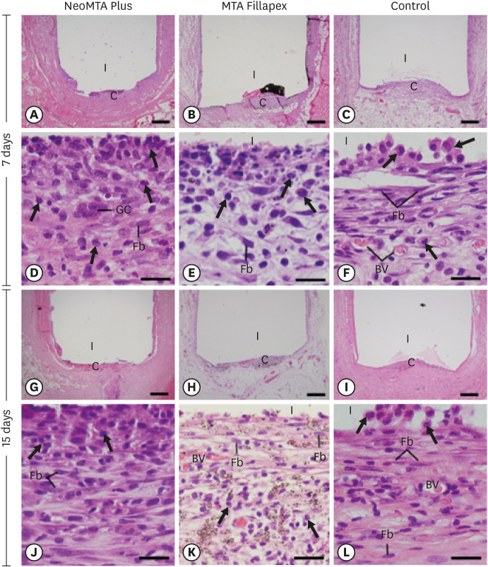
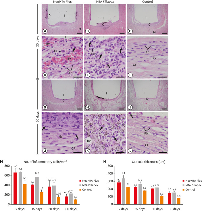
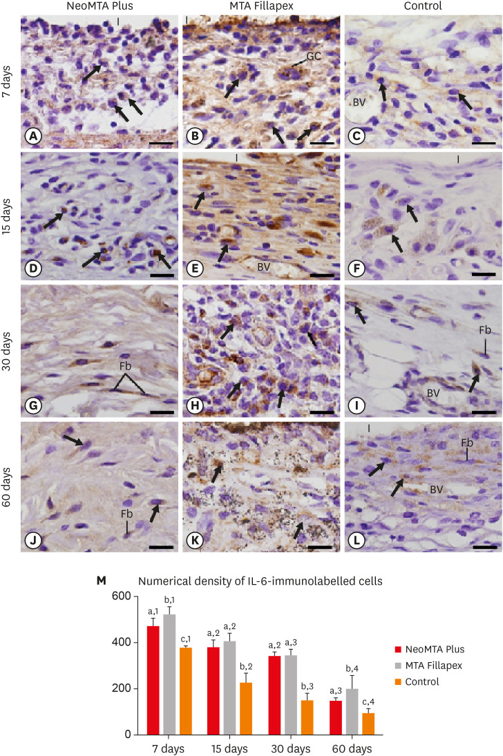
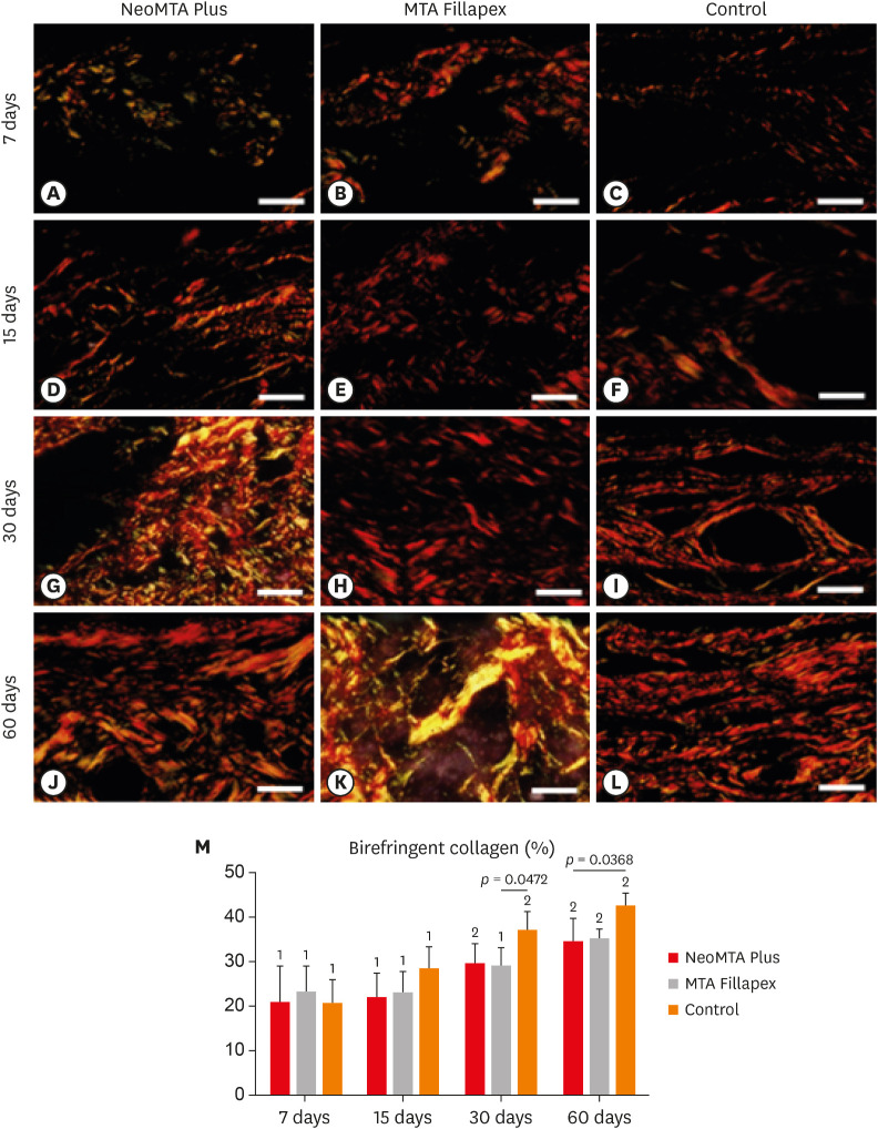
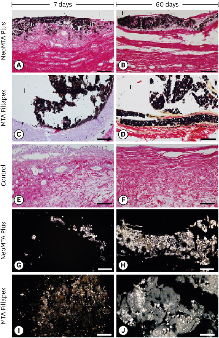
Materials and methods: Polyethylene tubes filled with NeoMTA Plus (n = 20), MTA Fillapex (n = 20), or nothing (control group, CG; n = 20) were inserted into the connective tissue in the dorsal subcutaneous layer of rats. After 7, 15, 30 and 60 days, the specimens were processed for paraffin embedding. The capsule thickness, collagen content, and number of inflammatory cells (ICs) and interleukin-6 (IL-6) immunolabeled cells were measured. von Kossa-positive structures were evaluated and unstained sections were analyzed under polarized light. Two-way analysis of variance was performed, followed by the post hoc Tukey test (p ≤ 0.05).
Results: At 7 days, the capsules around NeoMTA Plus and MTA Fillapex had more ICs and IL-6-immunostained cells than the CG. However, at 60 days, there was no significant difference in the IC number between NeoMTA Plus and the CG (p = 0.1137) or the MTA Fillapex group (p = 0.4062), although a greater number of IL-6-immunostained cells was observed in the MTA Fillapex group (p = 0.0353). From 7 to 60 days, the capsule thickness of the NeoMTA Plus and MTA Fillapex specimens significantly decreased, concomitantly with an increase in the collagen content. The capsules around root canal sealers showed positivity to the von Kossa stain and birefringent structures.
Conclusions: The NeoMTA Plus root canal sealer is biocompatible and exhibits bioactive potential.
Keywords: Bioactive potential; Biocompatibility; Immunohistochemistry; Inflammatory reaction; Interleukin-6.
Copyright © 2021. The Korean Academy of Conservative Dentistry.
Conflict of interest statement
Conflict of Interest: No potential conflict of interest relevant to this article was reported.
Figures
Similar articles
-
Reduced interleukin-6 immunoexpression and birefringent collagen formation indicate that MTA Plus and MTA Fillapex are biocompatible.Biomed Mater. 2018 Feb 20;13(3):035002. doi: 10.1088/1748-605X/aaa1f5. Biomed Mater. 2018. PMID: 29242419
-
Comparison of Bio-C Pulpo and MTA Repair HP with White MTA: effect on liver parameters and evaluation of biocompatibility and bioactivity in rats.Int Endod J. 2021 Sep;54(9):1597-1613. doi: 10.1111/iej.13567. Epub 2021 Jul 12. Int Endod J. 2021. PMID: 33999424
-
Biocompatibility and bioactive potential of NeoPUTTY calcium silicate-based cement: An in vivo study in rats.Int Endod J. 2024 Jun;57(6):713-726. doi: 10.1111/iej.14054. Epub 2024 Mar 11. Int Endod J. 2024. PMID: 38467586
-
Biocompatibility and Bioactive Potential of New Calcium Silicate-based Endodontic Sealers: Bio-C Sealer and Sealer Plus BC.J Endod. 2020 Oct;46(10):1470-1477. doi: 10.1016/j.joen.2020.07.011. Epub 2020 Jul 17. J Endod. 2020. PMID: 32682789
-
Cytotoxicity and biocompatibility of a new bioceramic endodontic sealer containing calcium hydroxide.Braz Oral Res. 2019;33:e042. doi: 10.1590/1807-3107bor-2019.vol33.0042. Epub 2019 May 16. Braz Oral Res. 2019. PMID: 31508725
Cited by
-
Bioactivity Potential of Bioceramic-Based Root Canal Sealers: A Scoping Review.Life (Basel). 2022 Nov 11;12(11):1853. doi: 10.3390/life12111853. Life (Basel). 2022. PMID: 36430988 Free PMC article.
-
An Updated Review on Properties and Indications of Calcium Silicate-Based Cements in Endodontic Therapy.Int J Dent. 2022 Oct 30;2022:6858088. doi: 10.1155/2022/6858088. eCollection 2022. Int J Dent. 2022. PMID: 36349079 Free PMC article. Review.
-
Sealing ability and microbial leakage of root-end filling materials: MTA versus epoxy resin: A systematic review and meta-analysis.Heliyon. 2021 Jul 7;7(7):e07494. doi: 10.1016/j.heliyon.2021.e07494. eCollection 2021 Jul. Heliyon. 2021. PMID: 34401555 Free PMC article. Review.
-
Calcium Silicate-Based Sealer Dentinal Tubule Penetration-A Systematic Review of In Vitro Studies.Materials (Basel). 2023 Mar 29;16(7):2734. doi: 10.3390/ma16072734. Materials (Basel). 2023. PMID: 37049028 Free PMC article. Review.
-
The osteoinductive potential of different root-end filling materials in a rat femur model.Sci Rep. 2024 Jan 24;14(1):2078. doi: 10.1038/s41598-024-52584-5. Sci Rep. 2024. PMID: 38267563 Free PMC article.
References
-
- Tomás-Catalá CJ, Collado-González M, García-Bernal D, Oñate-Sánchez RE, Forner L, Llena C, Lozano A, Moraleda JM, Rodríguez-Lozano FJ. Biocompatibility of new pulp-capping materials NeoMTA Plus, MTA Repair HP, and biodentine on human dental pulp stem cells. J Endod. 2018;44:126–132. - PubMed
-
- Cintra LTA, Benetti F, de Azevedo Queiroz ÍO, de Araújo Lopes JM, Penha de Oliveira SH, Sivieri Araújo G, Gomes-Filho JE. Cytotoxicity, biocompatibility, and biomineralization of the new high-plasticity MTA Material. J Endod. 2017;43:774–778. - PubMed
-
- Mondelli JAS, Hoshino RA, Weckwerth PH, Cerri PS, Leonardo RT, Guerreiro-Tanomaru JM, Tanomaru-Filho M, da Silva GF. Biocompatibility of mineral trioxide aggregate flow and biodentine. Int Endod J. 2019;52:193–200. - PubMed
-
- Parirokh M, Torabinejad M, Dummer PMH. Mineral trioxide aggregate and other bioactive endodontic cements: an updated overview - Part I: Vital pulp therapy. Int Endod J. 2018;51:177–205. - PubMed
-
- Siboni F, Taddei P, Prati C, Gandolfi MG. Properties of NeoMTA Plus and MTA Plus cements for endodontics. Int Endod J. 2017;50(Supplement 2):e83–e94. - PubMed
LinkOut - more resources
Full Text Sources
Other Literature Sources