

Potential Roles of Acute Phase Proteins in Cancer: Why Do Cancer Cells Produce or Take Up Exogenous Acute Phase Protein Alpha1-Antitrypsin?
- PMID: 33680966
- PMCID: PMC7933442
- DOI: 10.3389/fonc.2021.622076
Potential Roles of Acute Phase Proteins in Cancer: Why Do Cancer Cells Produce or Take Up Exogenous Acute Phase Protein Alpha1-Antitrypsin?
Abstract
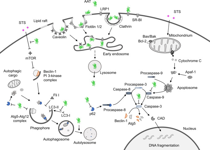
An association between acute-phase proteins (APPs) and cancer has long been established and there are numerous reports correlating altered levels and/or molecular forms of APPs with different types of cancers. Many authors have shown a positive correlation between high levels of APPs, like alpha1-antitrypsin (AAT), and unfavorable clinical outcome in cancers. Conversely, others proposed that high levels of APPs are probably just a part of nonspecific inflammatory response to cancer development. However, this might not be always true, because many cancerous cells produce or take up exogenous APPs. What is the biological significance of this and what benefit do cancer cells have from these proteins remains largely unknown. Recent data revealed that some APPs, including AAT, are able to enhance cancer cell resistance against anticancer drug-induced apoptosis and autophagy. In this review, we specifically discuss our own findings and controversies in the literature regarding the role of AAT in cancer.
Keywords: acute phase proteins; alpha1-antitrypsin; apoptosis; autophagy; cancer cells; cancer microenvironment.
Copyright © 2021 Janciauskiene, Wrenger, Günzel, Gründing, Golpon and Welte.
Conflict of interest statement
The authors declare that the research was conducted in the absence of any commercial or financial relationships that could be construed as a potential conflict of interest.
Figures
Similar articles
-
The role and importance of glycosylation of acute phase proteins with focus on alpha-1 antitrypsin in acute and chronic inflammatory conditions.J Proteome Res. 2014 Jul 3;13(7):3131-43. doi: 10.1021/pr500146y. Epub 2014 Jun 12. J Proteome Res. 2014. PMID: 24892502 Review.
-
Clinical Significance of SERPINA1 Gene and Its Encoded Alpha1-antitrypsin Protein in NSCLC.Cancers (Basel). 2019 Sep 4;11(9):1306. doi: 10.3390/cancers11091306. Cancers (Basel). 2019. PMID: 31487965 Free PMC article.
-
[Alpha-1 Antitrypsin Affects U0126-Induced Cytotoxicity in Colon Cancer Cell Line (HCT116)].Mol Biol (Mosk). 2016 Jan-Feb;50(1):174-8. doi: 10.7868/S0026898416010122. Mol Biol (Mosk). 2016. PMID: 27028823 Russian.
-
Functional protection by acute phase proteins alpha(1)-acid glycoprotein and alpha(1)-antitrypsin against ischemia/reperfusion injury by preventing apoptosis and inflammation.Circulation. 2000 Sep 19;102(12):1420-6. doi: 10.1161/01.cir.102.12.1420. Circulation. 2000. PMID: 10993862
-
Acute-phase protein α1-anti-trypsin: diverting injurious innate and adaptive immune responses from non-authentic threats.Clin Exp Immunol. 2015 Feb;179(2):161-72. doi: 10.1111/cei.12476. Clin Exp Immunol. 2015. PMID: 25351931 Free PMC article. Review.
Cited by
-
Plasma proteome fingerprint in kidney diseases.Front Mol Biosci. 2025 Jan 17;11:1494779. doi: 10.3389/fmolb.2024.1494779. eCollection 2024. Front Mol Biosci. 2025. PMID: 39896931 Free PMC article.
-
α‑1 Antitrypsin is a potential target of inflammation and immunomodulation (Review).Mol Med Rep. 2025 Apr;31(4):107. doi: 10.3892/mmr.2025.13472. Epub 2025 Feb 28. Mol Med Rep. 2025. PMID: 40017119 Free PMC article. Review.
-
Label-free quantitative proteomics reveals aberrant expression levels of LRG, C9, FN, A1AT and AGP1 in the plasma of patients with colorectal cancer.Clin Proteomics. 2023 Apr 6;20(1):15. doi: 10.1186/s12014-023-09407-y. Clin Proteomics. 2023. PMID: 37024778 Free PMC article.
-
Carbon-Based Nanomaterials as Drug Delivery Agents for Colorectal Cancer: Clinical Preface to Colorectal Cancer Citing Their Markers and Existing Theranostic Approaches.ACS Omega. 2023 Mar 14;8(12):10656-10668. doi: 10.1021/acsomega.2c06242. eCollection 2023 Mar 28. ACS Omega. 2023. PMID: 37008124 Free PMC article. Review.
-
Alu RNA induces NLRP3 expression through TLR7 activation in α-1-antitrypsin-deficient macrophages.JCI Insight. 2022 Jun 22;7(12):e158791. doi: 10.1172/jci.insight.158791. JCI Insight. 2022. PMID: 35730566 Free PMC article.
References
Publication types
LinkOut - more resources
Full Text Sources
Other Literature Sources
Research Materials
Miscellaneous