. 2021 Feb 18:9:614154.
doi: 10.3389/fchem.2021.614154. eCollection 2021.

Comparison of the Inhibitory Binding Modes Between the Planar Fascaplysin and Its Nonplanar Tetrahydro-β-carboline Analogs in CDK4

Affiliations

Comparison of the Inhibitory Binding Modes Between the Planar Fascaplysin and Its Nonplanar Tetrahydro-β-carboline Analogs in CDK4

Yan Liang et al. Front Chem. .

Abstract

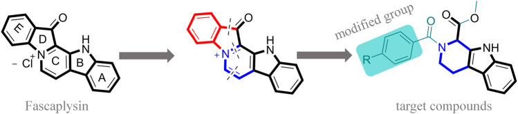
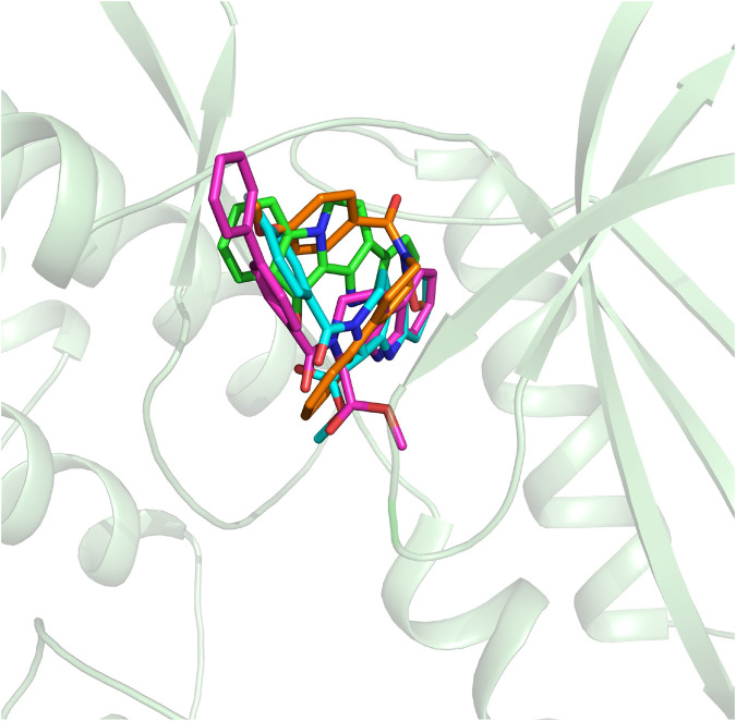
Fascaplysin is a natural marine product originating from sponges, attracting widespread attention due to its potential inhibitory activities against CDK4. However, its clinical application has been largely limited because of serious adverse effects caused by planar skeleton. To reduce the serious adverse effects, 18 tetrahydro-β-carboline analogs (compounds 6a-i and 7a-i) were designed and synthesized via breaking the planarity of fascaplysin, and the biological activities of the synthesized compounds were evaluated by MTT assay and CDK4/CycD3 enzyme inhibition assay. The title compounds showed varying degrees of inhibitory activities, especially the cytotoxicity of compound 6c against HeLa cells (IC50 = 1.03 ± 0.19 μM) with quite weak cytotoxicity toward the normal cells WI-38 (IC50 = 311.51 ± 56.06 μM), and the kinase inhibition test indicated that compound 6c was a potential CDK4 inhibitor. In order to further compare the action mechanisms of planar and nonplanar molecules on CDK4, the studied complexes of CDK4 bound with fascaplysin and three representative compounds (compound 6a-c) with bioactivities gradient were constructed by molecular docking and further verified through molecular dynamic simulation, which identified the key residues contributing to the ligands' binding. By comparing the binding modes of the constructed systems, it could be found that the residues contributing significantly to compound 6c's binding were highly consistent with those contributing significantly to fascaplysin's binding. Through the design, synthesis of the nonplanar fascaplysin derivatives, and binding mechanism analysis, some valuable hints for the discovery of antitumor drug candidates could be provided.

Keywords: MD simulation; MTT assay; fascaplysin; molecular docking; nonplanar.


Conflict of interest statement

The authors declare that the research was conducted in the absence of any commercial or financial relationships that could be construed as a potential conflict of interest.

Figures

FIGURE 1
FIGURE 1
Modification strategy of the target compounds.
FIGURE 2
FIGURE 2
Synthetic route of the target compounds.
FIGURE 3
FIGURE 3
Structural alignment of the initial docking poses of the studied molecules.
FIGURE 4
FIGURE 4
Root mean square deviations of protein backbone atoms, ligand heavy atoms, and binding site residue backbone atoms as a function of time in MD simulations.
FIGURE 5
FIGURE 5
Structural superimposition of the initial docking poses and the representative conformations of all the studied systems: (A) initial conformation (green) and representative snapshot (sky blue) of fascaplysin in CDK4; (B) initial conformation (cyan) and representative snapshot (gray) of compound 6a in CDK4; (C) initial conformation (orange) and representative snapshot (slate) of compound 6b in CDK4; (D) initial conformation (magentas) and representative snapshot (yellow) of compound 6c in CDK4.
FIGURE 6
FIGURE 6
Per-residue binding free energy decomposition of the residues with energy contribution: (A) fascaplysin-CDK4 system; (B) compound 6a-CDK4 system; (C) compound 6b-CDK4 system; (D) compound 6c-CDK4 system.
FIGURE 7
FIGURE 7
Key residues were identified by hierarchically clustering per‐residue energy contributions across four systems, and the residues were mainly divided into five Chains, namely Chain A–E. Residues favoring the binding of ligands were colored in red; residues hampering the binding of ligands were shown in blue (the highest one was colored as standard blue and the lower one was set to fade gradually to white); the white color here represented the residues with no energy contribution.
FIGURE 8
FIGURE 8
Comparison between the binding modes of the synthesized compounds and that of fascaplysin: (A) alignment of CDK4-compound 6a and CDK4-fascaplysin; (B) alignment of CDK4-compound 6b and CDK4-fascaplysin; (C) alignment of CDK4-compound 6c and CDK4-fascaplysin.
FIGURE 9
FIGURE 9
Comparison between the key residues consisting of the binding site of compound 6c and that of fascaplysin (A) fascaplysin in CDK4; (B) compound 6c in CDK4.

Similar articles

Cited by

References

    1. AMBER v. 16 (2016). San Francisco, CA: University of California.
    1. Bachurin S., Tkachenko S., Baskin I., Lermontova N., Mukhina T., Petrova L., et al. (2001). Neuroprotective and cognition-enhancing properties of MK-801 flexible analogs. Structure-activity relationships. Ann. N. Y. Acad. Sci. 939 (1), 219–236. 10.1111/j.1749-6632.2001.tb03629.x - DOI - PubMed
    1. Chen S., Guan X., Wang L. L., Li B., Sang X. B., Liu Y., et al. (2017). Fascaplysin inhibit ovarian cancer cell proliferation and metastasis through inhibiting CDK4. Gene 635, 3–8. 10.1016/j.gene.2017.09.009 - DOI - PubMed
    1. Choo J. R., Lee S. C. (2018). CDK4-6 inhibitors in breast cancer: current status and future development. Expet Opin. Drug Metabol. Toxicol. 14 (11), 1123–1138. 10.1080/17425255.2018.1541347 - DOI - PubMed
    1. Chotiner J. Y., Wolgemuth D. J., Wang P. J. (2019). Functions of cyclins and CDKs in mammalian gametogenesisdagger. Biol. Reprod. 101 (3), 591–601. 10.1093/biolre/ioz070 - DOI - PMC - PubMed

LinkOut - more resources