

Contributions of Embryonic HSC-Independent Hematopoiesis to Organogenesis and the Adult Hematopoietic System
- PMID: 33681211
- PMCID: PMC7930747
- DOI: 10.3389/fcell.2021.631699
Contributions of Embryonic HSC-Independent Hematopoiesis to Organogenesis and the Adult Hematopoietic System
Abstract
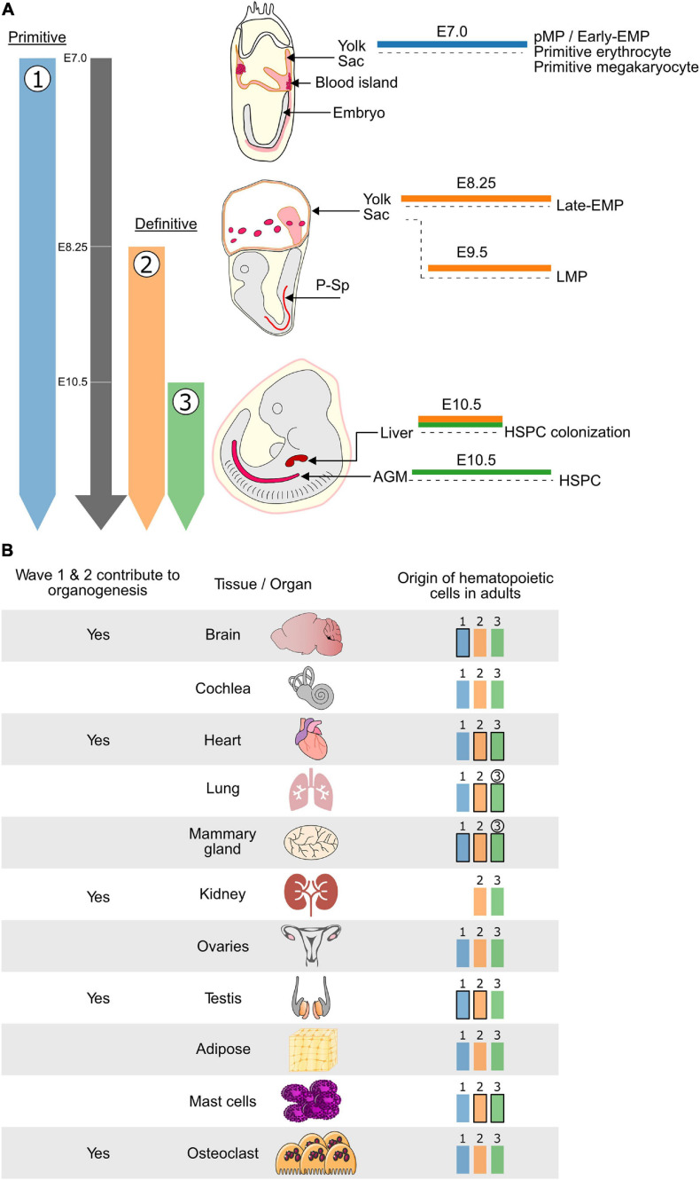
During ontogeny, the establishment of the hematopoietic system takes place in several phases, separated both in time and location. The process is initiated extra-embryonically in the yolk sac (YS) and concludes in the main arteries of the embryo with the formation of hematopoietic stem cells (HSC). Initially, it was thought that HSC-independent hematopoietic YS cells were transient, and only required to bridge the gap to HSC activity. However, in recent years it has become clear that these cells also contribute to embryonic organogenesis, including the emergence of HSCs. Furthermore, some of these early HSC-independent YS cells persist into adulthood as distinct hematopoietic populations. These previously unrecognized abilities of embryonic HSC-independent hematopoietic cells constitute a new field of interest. Here, we aim to provide a succinct overview of the current knowledge regarding the contribution of YS-derived hematopoietic cells to the development of the embryo and the adult hematopoietic system.
Keywords: HSC-independent hematopoiesis; embryonic hematopoiesis; hematopoietic waves; lineage tracing; macrophage; organogenesis; yolk sac.
Copyright © 2021 Neo, Lie-A-Ling, Fadlullah and Lacaud.
Conflict of interest statement
The authors declare that the research was conducted in the absence of any commercial or financial relationships that could be construed as a potential conflict of interest.
Figures
Similar articles
-
[Hematopoietic stem cell emergence and stem cell-independent hematopoiesis in the mouse embryo].Rinsho Ketsueki. 2018;59(7):915-921. doi: 10.11406/rinketsu.59.915. Rinsho Ketsueki. 2018. PMID: 30078803 Review. Japanese.
-
Hematopoietic stem cells in the mouse embryonic yolk sac.Stem Cells. 1996 May;14(3):269-80. doi: 10.1002/stem.140269. Stem Cells. 1996. PMID: 8724693 Review.
-
Early hematopoiesis and macrophage development.Semin Immunol. 2015 Dec;27(6):379-87. doi: 10.1016/j.smim.2016.03.013. Epub 2016 Mar 25. Semin Immunol. 2015. PMID: 27021646 Free PMC article. Review.
-
Mds1CreERT2, an inducible Cre allele specific to adult-repopulating hematopoietic stem cells.Cell Rep. 2021 Aug 17;36(7):109562. doi: 10.1016/j.celrep.2021.109562. Cell Rep. 2021. PMID: 34407416 Free PMC article.
-
Development of the hematopoietic system: expanding the concept of hematopoietic stem cell-independent hematopoiesis.Trends Cell Biol. 2024 Feb;34(2):161-172. doi: 10.1016/j.tcb.2023.06.007. Epub 2023 Jul 20. Trends Cell Biol. 2024. PMID: 37481335 Review.
Cited by
-
De novo hematopoiesis from the fetal lung.Blood Adv. 2023 Nov 28;7(22):6898-6912. doi: 10.1182/bloodadvances.2022008347. Blood Adv. 2023. PMID: 37729429 Free PMC article.
-
New insights into the endothelial origin of hematopoietic system inspired by "TIF" approaches.Blood Sci. 2024 Jul 16;6(4):e00199. doi: 10.1097/BS9.0000000000000199. eCollection 2024 Oct. Blood Sci. 2024. PMID: 39027902 Free PMC article. Review.
-
Integrative cross-species transcriptome analysis reveals earlier occurrence of myelopoiesis in pre-circulation primates compared to mice.Zool Res. 2024 Nov 18;45(6):1276-1286. doi: 10.24272/j.issn.2095-8137.2024.173. Zool Res. 2024. PMID: 39397246 Free PMC article.
-
Cannabidiol restores hematopoietic stem cell stemness in mouse through Atf2-Lrp6 axis after acute irradiation.MedComm (2020). 2025 Feb 13;6(2):e70092. doi: 10.1002/mco2.70092. eCollection 2025 Feb. MedComm (2020). 2025. PMID: 39949985 Free PMC article.
-
Immune modulatory stem cells represent a significant component of the immune system.Front Immunol. 2025 Mar 3;16:1543495. doi: 10.3389/fimmu.2025.1543495. eCollection 2025. Front Immunol. 2025. PMID: 40098974 Free PMC article. No abstract available.
References
Publication types
LinkOut - more resources
Full Text Sources
Other Literature Sources
Miscellaneous