

Evaluation of CD44 and TGF-B Expression in Oral Carcinogenesis
- PMID: 33681421
- PMCID: PMC7921764
- DOI: 10.30476/DENTJODS.2020.84393.1079
Evaluation of CD44 and TGF-B Expression in Oral Carcinogenesis
Abstract
Statement of the problem: Oral squamous cell carcinoma (OSCC) is the most common malignancy of the oral cavity. Early diagnosis of OSCC by using biomarkers provides preventive treatment approach to suppress the disease in early stages. CD44 as a cancer stem cell (CSC) marker may be cleaved by MT1-MMP and plays an important role in migration of cancer cells. TGF-B promotes formation of invasive cancer cells phenotype through epithelial mesenchymal transition (EMT) and induces MT1-MMP formation.
Purpose: The aim of this study was to evaluate the expression of TGF-B and CD44 in leukoplakia (premalignant lesion), squamous cell carcinoma (SCC), and normal oral mucosa to determine the role of these markers in the carcinogenesis process of the oral mucosa.
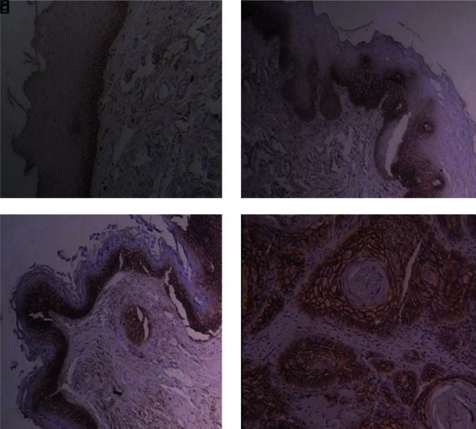
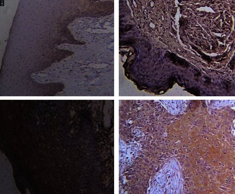
Materials and method: In this retrospective study, the expression of TGF-B and CD44 were evaluated in 55 paraffin-embedded specimens (10normal mucosa, 15 non-dysplastic leukoplakia, 15 dysplastic leukoplakia, and 15 OSCC) by immunohistochemistry. Statistical analyses were performed using Kruskal-Wallis, Mann-Whitney, and Spearman's rank correlation tests.
Results: Evaluation of CD44 and TGF-B expression in the four studied groups showed statistical significant difference for each marker (p< 0.001). Pairwise comparison of CD44 and TGF-B expression in all groups except normal mucosa and non-dysplastic leukoplakia demonstrated statistical significant difference. In addition, there was positive significant correlation between two markers (r= 0.914, p< 0.001). Diagnostic test's accuracy for identification of OSCC and dysplastic leukoplakia from non-dysplastic leukoplakia and normal tissues and recognition of OSCC from dysplastic leukoplakia showed optimum sensitivity and specificity.
Conclusion: Increased expression of CD44 as a cancer stem cell marker and TGF-B as an EMT marker from normal mucosa to non-dysplastic leukoplakia, dysplastic leukoplakia, and OSCC and also the significant correlation between these two markers indicated their role in carcinogenesis of oral mucosa.
Keywords: CD44; Dysplasia; Oral squamous cell carcinoma; TGF-B.
Copyright: © Journal of Dentistry.
Figures
Similar articles
-
Immunohistochemical Expression of Differentiated Embryonic Chondrocyte 1 and Cluster of Differentiation 44 in Oral Potentially Malignant Disorders.Medicina (Kaunas). 2025 Feb 1;61(2):251. doi: 10.3390/medicina61020251. Medicina (Kaunas). 2025. PMID: 40005368 Free PMC article.
-
New insights into the role of the oral leukoplakia microenvironment in malignant transformation.Front Oral Health. 2024 Feb 21;5:1363052. doi: 10.3389/froh.2024.1363052. eCollection 2024. Front Oral Health. 2024. PMID: 38450102 Free PMC article. Review.
-
Evaluation of CD24 and CD44 as cancer stem cell markers in squamous cell carcinoma and epithelial dysplasia of the oral cavity by q- RT-PCR.Dent Res J (Isfahan). 2020 May 23;17(3):208-212. eCollection 2020 May-Jun. Dent Res J (Isfahan). 2020. PMID: 32774798 Free PMC article.
-
Preliminary Study of the Cancer Stem Cells' Biomarker CD147 in Leukoplakia: Dysplasia and Squamous Cell Carcinoma of Oral Epithelial Origin.Cureus. 2023 May 9;15(5):e38807. doi: 10.7759/cureus.38807. eCollection 2023 May. Cureus. 2023. PMID: 37303447 Free PMC article.
-
Influence of site and smoking on malignant transformation in the oral cavity: Is the microbiome the missing link?Front Oral Health. 2023 Mar 23;4:1166037. doi: 10.3389/froh.2023.1166037. eCollection 2023. Front Oral Health. 2023. PMID: 37035251 Free PMC article. Review.
Cited by
-
Can precancerous stem cells be risk markers for malignant transformation in the oral mucosa?Cell Mol Biol Lett. 2023 Apr 7;28(1):30. doi: 10.1186/s11658-023-00441-0. Cell Mol Biol Lett. 2023. PMID: 37029348 Free PMC article.
-
HIV-1 Tat-induced disruption of epithelial junctions and epithelial-mesenchymal transition of oral and genital epithelial cells lead to increased invasiveness of neoplastic cells and the spread of herpes simplex virus and cytomegalovirus.Front Immunol. 2025 Feb 13;16:1541532. doi: 10.3389/fimmu.2025.1541532. eCollection 2025. Front Immunol. 2025. PMID: 40018040 Free PMC article. Review.
-
Immunohistochemical Expression of Differentiated Embryonic Chondrocyte 1 and Cluster of Differentiation 44 in Oral Potentially Malignant Disorders.Medicina (Kaunas). 2025 Feb 1;61(2):251. doi: 10.3390/medicina61020251. Medicina (Kaunas). 2025. PMID: 40005368 Free PMC article.
-
New insights into the role of the oral leukoplakia microenvironment in malignant transformation.Front Oral Health. 2024 Feb 21;5:1363052. doi: 10.3389/froh.2024.1363052. eCollection 2024. Front Oral Health. 2024. PMID: 38450102 Free PMC article. Review.
-
Immunoexpression of CD44, p16 and VEGF in oral cancer.J Oral Maxillofac Pathol. 2024 Apr-Jun;28(2):253-260. doi: 10.4103/jomfp.jomfp_195_23. Epub 2024 Jul 11. J Oral Maxillofac Pathol. 2024. PMID: 39157839 Free PMC article.
References
-
- Neville B, Damm DD, Allen C, Chi A. Oral and maxillofacial pathology. 4th ed. WB Saunders, Elsevier: Missouri; 2016. p.928.
-
- Oliveira LR, Castilho-Fernandes A, Oliveira-Costa JP, Soares FA, Zucoloto S, Ribeiro-Silva A. CD44+/CD133+ immunophenotype and matrix metalloproteinase-9: Influence on prognosis in early-stage oral squamous cell carcinoma. Head Neck. 2014; 36: 1718–1726. - PubMed
-
- Saghravanian N, Anvari K, Ghazi N, Memar B, Shahsavari M, Aghaee MA. Expression of p63 and CD44 in oral squamous cell carcinoma and correlation with clinicopathological parameters. Arch Oral Biol. 2017; 82: 160–165. - PubMed
-
- Godge PY, Poonja LS. Quantitative assessment of expression of cell adhesion molecule (CD44) splice variants: CD44 standard (CD44s) and v5, v6 isoforms in oral leukoplakias: an immunohistochemical study. Indian J Dent Res. 2011; 22: 493–494. - PubMed
-
- Salehinejad J, Sharifi N, Amirchaghmaghi M, Ghazi N, Shakeri MT, Ghazi A. Immunohistochemical expression of p16 protein in oral squamous cell carcinoma and lichen planus. Annals of Diagnostic Pathology. 2014; 18: 210–213. - PubMed
LinkOut - more resources
Full Text Sources
Research Materials
Miscellaneous