

Acellular dermal matrix in skin wound healing in rabbits - histological and histomorphometric analyses
- PMID: 33681941
- PMCID: PMC7920408
- DOI: 10.6061/clinics/2021/e2066
Acellular dermal matrix in skin wound healing in rabbits - histological and histomorphometric analyses
Abstract
Objectives: To analyze the histology and histomorphometry of healing associated with acellular dermal matrix in skin wounds in rabbits.
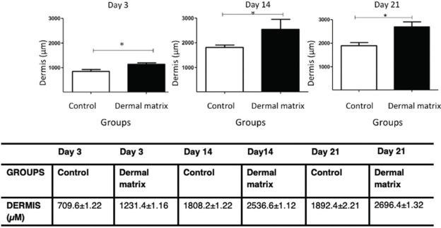
Methods: Twelve male rabbits were divided into two groups: the control group (CG) and the matrix group (MG). Three skin wounds with a total area of 20 × 20 mm were created on the dorsal region of each animal. Photographic records of the lesions taken over a 21-day period and use of the ImageJ program allowed calculation of the wound contraction rate. The lesions were biopsied on days 3, 14 and 21 for histomorphometric analysis to define the thicknesses of the dermis and epidermis (hematoxylin-eosin) and calculate the densities of type I and type III collagen (picrosirius).
Results: No significant difference in the healing rate was found between the groups (p>0.05). The MG presented greater epidermal thickness on day 3 (p<0.05) and on days 14 and 21 (p<0.001). The MG presented greater dermal thickness throughout the study period (p<0.05). The type I collagen density was higher in the MG throughout the study period (p<0.05), and the type III collagen density was higher in the MG on days 3 and 14 (p<0.05) and on day 21 (p<0.001).
Conclusion: The use of acellular dermal matrix increased the thickness of the dermal and epidermal layers and the amount of type I and III collagen during skin wound healing and did not alter the rate of wound contraction.
Conflict of interest statement
No potential conflict of interest was reported.
Figures


Similar articles
-
[Effects on collagen I and III after transplantation of porcine acellular dermal matrix with hyaluronic acid].Zhonghua Yi Xue Za Zhi. 2011 May 17;91(18):1276-80. Zhonghua Yi Xue Za Zhi. 2011. PMID: 21756802 Chinese.
-
[Influence of porcine urinary bladder matrix and porcine acellular dermal matrix on wound healing of full-thickness skin defect in diabetic mice].Zhonghua Shao Shang Za Zhi. 2020 Dec 20;36(12):1130-1138. doi: 10.3760/cma.j.cn501120-20200901-00399. Zhonghua Shao Shang Za Zhi. 2020. PMID: 33379849 Chinese.
-
Influence of hyaluronic acid on wound healing using composite porcine acellular dermal matrix grafts and autologous skin in rabbits.Int Wound J. 2013 Oct;10(5):562-72. doi: 10.1111/j.1742-481X.2012.01023.x. Epub 2012 Jun 11. Int Wound J. 2013. PMID: 22682212 Free PMC article.
-
Human acellular dermal wound matrix for treatment of DFU: literature review and analysis.J Wound Care. 2015 Mar;24(3):128; 129-34. doi: 10.12968/jowc.2015.24.3.128. J Wound Care. 2015. PMID: 25764957 Review.
-
Effect of collagen matrices on dermal wound healing.Adv Drug Deliv Rev. 2003 Nov 28;55(12):1595-611. doi: 10.1016/j.addr.2003.08.003. Adv Drug Deliv Rev. 2003. PMID: 14623403 Review.
Cited by
-
Characterization and Strength Quality of the Oryctolagus cuniculus Leather Compared to Oreochromis niloticus Leather.ScientificWorldJournal. 2022 Oct 14;2022:4561404. doi: 10.1155/2022/4561404. eCollection 2022. ScientificWorldJournal. 2022. PMID: 36277128 Free PMC article.
-
A long-term follow-up study of diabetic foot ulcer using micronized acellular dermal matrix.Int Wound J. 2023 May;20(5):1622-1637. doi: 10.1111/iwj.14018. Epub 2022 Nov 15. Int Wound J. 2023. PMID: 36377547 Free PMC article.
-
Wound Healing after Acellular Dermal Substitute Positioning in Dermato-Oncological Surgery: A Prospective Comparative Study.Life (Basel). 2023 Feb 7;13(2):463. doi: 10.3390/life13020463. Life (Basel). 2023. PMID: 36836820 Free PMC article.
-
The Effect of Fetal Bovine Acellular Dermal Matrix Seeded with Wharton's Jelly Mesenchymal Stem Cells for Healing Full-Thickness Skin Wounds.Genes (Basel). 2023 Apr 13;14(4):909. doi: 10.3390/genes14040909. Genes (Basel). 2023. PMID: 37107668 Free PMC article.
-
Effects of Laurus nobilis Leaf Extract on Healing of Experimentally Induced Wounds in Rabbits.Vet Med Int. 2024 Oct 15;2024:2889480. doi: 10.1155/2024/2889480. eCollection 2024. Vet Med Int. 2024. PMID: 39444801 Free PMC article.
References
-
- Young A, McNaught CE. The physiology of wound healing. Surgery (Oxford) 2011;29(10):475–9.
MeSH terms
Substances
LinkOut - more resources
Full Text Sources
Other Literature Sources
