. 2021 Mar 8:76:e2484.
doi: 10.6061/clinics/2021/e2484. eCollection 2021.

miR-139-5p protects septic mice with acute lung injury by inhibiting Toll-like receptor 4/Myeloid differentiation factor 88/Nuclear factor-&mac_kgr;B signaling pathway

Affiliations

miR-139-5p protects septic mice with acute lung injury by inhibiting Toll-like receptor 4/Myeloid differentiation factor 88/Nuclear factor-&mac_kgr;B signaling pathway

Xiuxiu Zhang et al. Clinics (Sao Paulo). .

Abstract

Objectives: To investigate the role of miR-139-5p and the TLR4/MyD88/NF-κB signaling pathway in acute lung injury in septic mice.

Method: A total of 140 healthy male SPF C57BL/6 mice were divided into seven groups, i.e., Normal, Control, NC, miR-139-5p mimic, miR-139-5p inhibitor, TAK-242, and miR-139-5p inhibitor+TAK-242 groups. The levels of miR-139-5p, proteins related to the TLR4/MyD88/NF-κB signaling pathway (TLR4, MyD88, and p-NF-κB p50), and MPO, SOD, GSH, and MDA in lung tissue were measured. The lung tissue wet-to-dry mass ratio (W/D), arterial oxygen partial pressure (PaO2), and carbon dioxide partial pressure (PaCO2) were measured.

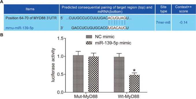
Results: A web-based bioinformatic tool predicted that MyD88 was a target of miR-139-5p, which was verified by a dual luciferase reporter assay. Compared with those in the Normal group, the levels of miR-139-5p, PaO2, SOD, and GSH were significantly lower, while those of TLR4, MyD88, p-NF-κB p50, W/D, PaCO2, IL-1β, TNF-α, IL-6, MPO, and MDA were higher in all other groups. Moreover, compared with their levels in the Control group, these indicators exhibited contrasting results in the miR-139-5p mimic and TAK-242 groups, but were similar in the miR-139-5p inhibitor group. In the miR-139-5p inhibitor+TAK-242 group, acute lung injury, aggravated by miR-139-5p inhibitor, was partially rescued by TAK-242.

Conclusion: miR-139-5p inhibits the TLR4/MyD88/NF-κB signaling pathway to alleviate acute lung injury in septic mice.


Conflict of interest statement

No potential conflict of interest was reported.

Figures

Figure 1
Figure 1. miR-139-5p targets MyD88 gene expression. The 3'-UTR region in which miR-139-5p binds to MyD88 (A); results of the dual luciferase reporter assay (B). The asterisk (*) indicates a significant difference compared with the NC group, where p<0.05. MyD88, myeloid differentiation factor 88.
Figure 2
Figure 2. Expression of miR-139-5p, TLR4, MyD88, and NF-κB p50 in the lung tissue of each group. The mRNA levels of miR-139-5p, TLR4, MyD88, and NF-κB p50 in mouse lung tissue (A); The protein expression levels of TLR4, MyD88, NF-κB p50 and p-NF-κB p50 in mouse lung tissue (B-C). Significant comparisons with each group, where p<0.05, are indicated by: “*” for the Normal group, “#” for the Control group, “%” for NC group, “&” for the miR-139-5p mimic group, “$” for the miR-139-5p inhibitor group, and “@” for the TAK-242 group. TLR4, toll-like receptor 4; MyD88, myeloid differentiation factor 88.
Figure 3
Figure 3. The W/D, PaO2 and PaCO2 of mice in each group. W/D of mouse lung tissue (A); PaO2 in mice (B); PaCO2 in mice (C). Significant comparisons with each group, where p<0.05, are indicated by: “*” for the Normal group, “#” for the Control group, “%” for NC group, “&” for the miR-139-5p mimic group, “$” for the miR-139-5p inhibitor group, and “@” for the TAK-242 g roup PaO2, arterial oxygen partial pressure; W/D, lung tissue wet-to-dry mass ratio; PaCO2, carbon dioxide partial pressure.
Figure 4
Figure 4. Serum levels of the inflammatory factors, IL-1β, IL-6, and TNF-α in mice of each group. IL-1β levels (A); IL-6 levels (B); TNF-α levels (C). Significant comparisons with each group, where p<0.05, are indicated by: “*” for the Normal group, “#” for the Control group, “%” for NC group, “&” for the miR-139-5p mimic group, “$” for the miR-139-5p inhibitor group, and “@” for the TAK-242 group.
Figure 5
Figure 5. Levels of MPO (A), SOD (B), GSH (C), MDA (D) in the lung tissue of mice in each group. Significant comparisons with each group, where p<0.05, are indicated by: “*” for the Normal group, “#” for the Control group, “%” for NC group, “&” for the miR-139-5p mimic group, “$” for the miR-139-5p inhibitor group, and “@” for the TAK-242 group. MPO, myeloperoxidase; SOD, superoxide dismutase; GSH, glutathione; MDA, malondialdehyde.

Similar articles

Cited by

References

    1. Liu W, Liu K, Zhang S, Shan L, Tang J. Tetramethylpyrazine Showed Therapeutic Effects on Sepsis-Induced Acute Lung Injury in Rats by Inhibiting Endoplasmic Reticulum Stress Protein Kinase RNA-Like Endoplasmic Reticulum Kinase (PERK) Signaling-Induced Apoptosis of Pulmonary Microvascular Endothelial Cells. Med Sci Monit. 2018;24:1225–31. doi: 10.12659/MSM.908616. - DOI - PMC - PubMed
    1. Park I, Kim M, Choe K, Song E, Seo H, Hwang Y, et al. Neutrophils disturb pulmonary microcirculation in sepsis-induced acute lung injury. Eur Respir J. 2019;53(3):1800786. doi: 10.1183/13993003.00786-2018. - DOI - PMC - PubMed
    1. Xue M, Sun Z, Shao M, Yin J, Deng Z, Zhang J, et al. Diagnostic and prognostic utility of tissue factor for severe sepsis and sepsis-induced acute lung injury. J Transl Med. 2015;13:172. doi: 10.1186/s12967-015-0518-9. - DOI - PMC - PubMed
    1. Sun Y, Sun L, Liu S, Song J, Cheng J, Liu J. Effect of emodin on Aquaporin 5 expression in rats with sepsis-induced acute lung injury. J Tradit Chin Med. 2015;35(6):679–84. doi: 10.1016/S0254-6272(15)30159-X. - DOI - PubMed
    1. Mo Y, Lou Y, Zhang A, Zhang J, Zhu C, Zheng B, et al. PICK1 Deficiency Induces Autophagy Dysfunction via Lysosomal Impairment and Amplifies Sepsis-Induced Acute Lung Injury. Mediators Inflamm. 2018;2018:6757368. doi: 10.1155/2018/6757368. - DOI - PMC - PubMed