

Cell death as part of innate immunity: Cause or consequence?
- PMID: 33682112
- PMCID: PMC8274179
- DOI: 10.1111/imm.13325
Cell death as part of innate immunity: Cause or consequence?
Abstract
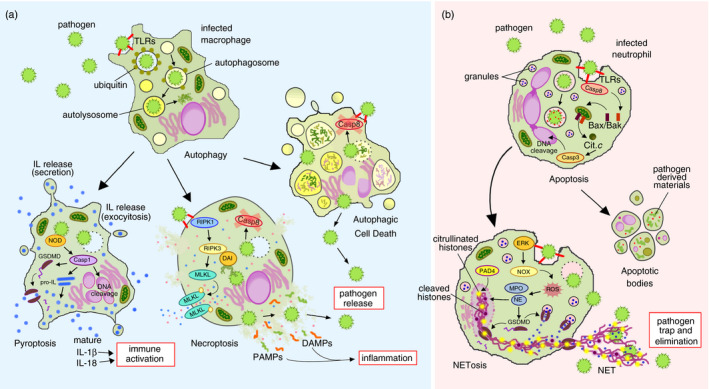
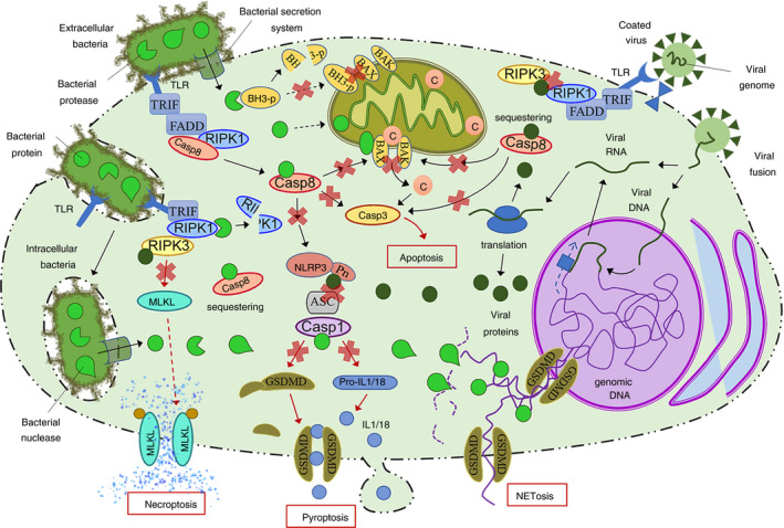
Regulated or programmed cell death plays a critical role in the development and tissue organization and function. In addition, it is intrinsically connected with immunity and host defence. An increasing cellular and molecular findings cause a change in the concept of cell death, revealing an expanding network of regulated cell death modalities and their biochemical programmes. Likewise, recent evidences demonstrate the interconnection between cell death pathways and how they are involved in different immune mechanisms. This work provides an overview of the main cell death programmes and their implication in innate immunity not only as an immunogenic/inflammatory process, but also as an active defence strategy during immune response and at the same time as a regulatory mechanism.
Keywords: apoptosis; innate immunity; molecular pathways; necrosis; programmed cell death; regulated.
© 2021 John Wiley & Sons Ltd.
Conflict of interest statement
There are not conflicts of interest associated with this work.
Figures
Similar articles
-
Multiple roles of caspase-8 in cell death, inflammation, and innate immunity.J Leukoc Biol. 2021 Jan;109(1):121-141. doi: 10.1002/JLB.3MR0420-305R. Epub 2020 Jun 12. J Leukoc Biol. 2021. PMID: 32531842 Free PMC article. Review.
-
Innate Immunity as an Executor of the Programmed Death of Individual Organisms for the Benefit of the Entire Population.Int J Mol Sci. 2021 Dec 15;22(24):13480. doi: 10.3390/ijms222413480. Int J Mol Sci. 2021. PMID: 34948277 Free PMC article. Review.
-
Non-apoptotic functions of cell death effectors in inflammation and innate immunity.Microbes Infect. 2012 Nov;14(14):1241-53. doi: 10.1016/j.micinf.2012.06.005. Epub 2012 Jun 21. Microbes Infect. 2012. PMID: 22728757 Review.
-
Programmed cell death as a defence against infection.Nat Rev Immunol. 2017 Mar;17(3):151-164. doi: 10.1038/nri.2016.147. Epub 2017 Jan 31. Nat Rev Immunol. 2017. PMID: 28138137 Free PMC article. Review.
-
Cell death in chronic inflammation: breaking the cycle to treat rheumatic disease.Nat Rev Rheumatol. 2020 Sep;16(9):496-513. doi: 10.1038/s41584-020-0455-8. Epub 2020 Jul 8. Nat Rev Rheumatol. 2020. PMID: 32641743 Review.
Cited by
-
The Role of Programmed Necrosis in Colorectal Cancer.Cancers (Basel). 2022 Sep 1;14(17):4295. doi: 10.3390/cancers14174295. Cancers (Basel). 2022. PMID: 36077828 Free PMC article. Review.
-
DEAD/H-Box Helicases in Immunity, Inflammation, Cell Differentiation, and Cell Death and Disease.Cells. 2022 May 11;11(10):1608. doi: 10.3390/cells11101608. Cells. 2022. PMID: 35626643 Free PMC article. Review.
-
Into the storm: the imbalance in the yin-yang immune response as the commonality of cytokine storm syndromes.Front Immunol. 2024 Sep 10;15:1448201. doi: 10.3389/fimmu.2024.1448201. eCollection 2024. Front Immunol. 2024. PMID: 39318634 Free PMC article. Review.
-
A deadly taste: linking bitter taste receptors and apoptosis.Apoptosis. 2025 Apr;30(3-4):674-692. doi: 10.1007/s10495-025-02091-3. Epub 2025 Feb 20. Apoptosis. 2025. PMID: 39979526 Free PMC article. Review.
-
Infection of Galleria mellonella (Lepidoptera) Larvae With the Entomopathogenic Fungus Conidiobolus coronatus (Entomophthorales) Induces Apoptosis of Hemocytes and Affects the Concentration of Eicosanoids in the Hemolymph.Front Physiol. 2022 Jan 6;12:774086. doi: 10.3389/fphys.2021.774086. eCollection 2021. Front Physiol. 2022. PMID: 35069239 Free PMC article.
References
-
- Allam R, Kumar SV, Darisipudi MN, Anders H‐J. Extracellular histones in tissue injury and inflammation. J Mol Med. 2014;92:465–72. - PubMed
-
- Godlewski M, Kobylińska A. Programmed cell death‐strategy for maintenance cellular organisms homeostasis. Postepy Hig Med Dosw(Online). 2016;70:1229. - PubMed
-
- Linkermann A, Stockwell BR, Krautwald S, Anders H‐J. Regulated cell death and inflammation: an auto‐amplification loop causes organ failure. Nat Rev Immunol. 2014;14:759–67. - PubMed
-
- Pasparakis M, Vandenabeele P. Necroptosis and its role in inflammation. Nature. 2015;517:311–20. - PubMed
Publication types
MeSH terms
LinkOut - more resources
Full Text Sources
Other Literature Sources
