. 2021 Aug;288(16):4833-4848.
doi: 10.1111/febs.15802. Epub 2021 Mar 26.

Reg1 and Snf1 regulate stress-induced relocalization of protein phosphatase-1 to cytoplasmic granules

Affiliations

Reg1 and Snf1 regulate stress-induced relocalization of protein phosphatase-1 to cytoplasmic granules

Helena Maria Schnell et al. FEBS J. 2021 Aug.

Abstract

The compartmentalization of cellular function is achieved largely through the existence of membrane-bound organelles. However, recent work suggests a novel mechanism of compartmentalization mediated by membraneless structures that have liquid droplet-like properties and arise through phase separation. Cytoplasmic stress granules (SGs) are the best characterized and are induced by various stressors including arsenite, heat shock, and glucose deprivation. Current models suggest that SGs play an important role in protein homeostasis by mediating reversible translation attenuation. Protein phosphatase-1 (PP1) is a central cellular regulator responsible for most serine/threonine dephosphorylation. Here, we show that upon arsenite stress, PP1's catalytic subunit Glc7 relocalizes to punctate cytoplasmic granules. This altered localization requires PP1's recently described maturation pathway mediated by the multifunctional ATPase Cdc48 and PP1's regulatory subunit Ypi1. Glc7 relocalization is mediated by its regulatory subunit Reg1 and its target Snf1, the AMP-dependent protein kinase. Surprisingly, Glc7 granules are highly specific to arsenite and appear distinct from canonical SGs. Arsenite induces potent translational inhibition, and translational recovery is strongly dependent on Glc7, but independent of Glc7's well-established role in regulating eIF2α. These results suggest a novel form of stress-induced cytoplasmic granule and a new mode of translational control by Glc7.

Keywords: Glc7; Reg1; Snf1; protein phosphatase-1; stress granules.


Conflict of interest statement

Conflicts of Interest: The authors declare that they have no conflict of interest.

Figures
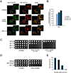
Fig. 1.
Fig. 1.
Glc7 localizes to arsenite-induced cytoplasmic granules. (A) Fluorescence microscopy of Glc7-mCherry and Hsp104-yeGFP after treatment with sodium arsenite (1 mM for 2 hours) and subsequent wash out (3 hours). Scale bar, 10 μm. (B) Relative protein abundance of Glc7 in response to sodium arsenite (1 mM) as determined by TMT-based mass spectrometry (raw proteomic data from [23]). Error bars represent standard deviations from biologic triplicates. (C) Sensitivity of wild-type and hypomorphic glc7 mutants to sodium arsenite (concentrations as indicated). Cells were spotted in 3-fold serial dilutions and cultured at 30°C for 2–4 days. Similar results were obtained in more than ten (panel A) and two (panel C) independent experiments. (D) Wild-type resistance of the Glc7-mCherry strain to sodium arsenite (0.25 mM). Cells were spotted in 3-fold serial dilutions and cultured at 30°C for 2–3 days. E) Time dependence of resolution of cytoplasmic foci after arsenite wash-out. Cells were treated with sodium arsenite (1 mM for 2 hours) which was then washed out and cytoplasmic foci were counted at the indicated time points. Cells were counted in three groups of 100, and errors bars reflect standard deviations.
Fig. 2.
Fig. 2.
Cdc48 and Ypi1 are required for Glc7 localization to arsenite-induced granules. (A) Fluorescence microscopy of Glc7-mCherry and Hsp104-yeGFP in wild-type and cdc48-ts strains after treatment with sodium arsenite (1 mM for 2 hours). Scale bar, 10 μm. (B) Knockdown of Ypi1 protein levels using the pGAL1–3xHA-YPI1 strain. Whole cell extracts were prepared at the indicated time points and analyzed by SDS-PAGE followed by immunoblotting. Upper panel, anti-HA antibody; lower panel, anti-Rpn5 antibody (loading control). (C) Fluorescence microscopy of Glc7-yeGFP (upper panels) in wild-type and pGAL1–3xHA-YPI1 yeast cultured in either galactose or glucose containing media followed by treatment with sodium arsenite (1 mM for 2 hours). The same experiment was performed with Hsp104-GFP in wild-type and pGAL1–3xHA-YPI1 yeast (lower panels). Scale bar, 10 μm. (D) Relative protein abundance of Ypi1 and GAPDH (control protein) in response to sodium arsenite (1 mM) as determined by TMT-based mass spectrometry (raw proteomic data from [23]). Error bars represent standard deviations from biologic triplicates. (E) Schematic representation of the PACE motif with the distance to YPI1 and RPN11 start sites indicated. (F) YPI1 transcription in wild-type and rpn4Δ cells in response to sodium arsenite (1 mM for 2 hours) as determined by RT-PCR. ACT1 mRNA serves as a control. Similar results were obtained in at least two independent experiments for all panels (except D).
Fig. 3.
Fig. 3.
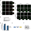
Localization of Glc7 to arsenite-induced granules is regulated by Reg1 and Snf1. (A) Fluorescence microscopy of Glc7-mCherry and Hsp104-yeGFP after treatment of wild-type, reg1Δ, and snf1Δreg1Δ cells with sodium arsenite (1 mM for 2 hours). Scale bar, 10 μm. (B) Quantitation of fluorescence intensity of Glc7-mCherry and Hsp104-GFP from individual cells. Graphs show fluorescence intensity along the lines shown in the left-hand panels. (C) Quantitation of the percentage of cells showing substantial co-localization of Glc7 and Hsp104. For each strain and condition, 150 consecutive cells were counted in groups of 50. Error bars reflect standard deviations. (D) Growth of Glc7 cofactor null mutants upon exposure to sodium arsenite (1.25 mM). Cells were spotted in 3-fold serial dilutions and cultured at 30°C for 2–4 days. (E) Localization of GFP-Reg1 in response to sodium arsenite (1 mM for 2 hours). Scale bar, 10 μm. (F) Fluorescence microscopy of Glc7-mCherry and Hsp104-yeGFP after treatment of wild-type, reg1Δ, and snf1-T210A reg1Δ cells with sodium arsenite (1 mM for 2 hours). Scale bar, 10 μm. (G) Growth of wild-type, reg1Δ, and snf1Δreg1Δ strains upon exposure to sodium arsenite (0.8 mM). Cells were spotted in 3-fold serial dilutions and cultured at 30°C for 2–4 days. Similar results were obtained in at least two independent experiments (panels A-G); the failure of Glc7 to form foci in the reg1Δ mutant was seen in more than ten independent experiments.
Fig. 4.
Fig. 4.
Snf1 co-factors required for Glc7 localization to arsenite-induced granules. Fluorescence microscopy of Glc7-mCherry and Hsp104-yeGFP after treatment of wild-type, reg1Δ, snf4Δreg1Δ, gal83Δsip1Δreg1Δ, gal83Δsip2Δreg1Δ, and sip1Δsip2Δreg1Δ yeast with sodium arsenite (1 mM for 2 hours). This is a composite panel in which some mutants were examined in separate experiments on different days. However, each experiment was performed with its own untreated and arsenite-treated wild-type and reg1Δ controls. Similar results were obtained in two independent experiments; see also Fig. 3A,F. Scale bars, 10 μm.
Fig. 5.
Fig. 5.
Arsenite-induced cytoplasmic granules are distinct from stress granules. (A) Fluorescence microscopy showing Glc7-mCherry and Hsp104-yeGFP after treatment with various stressors (1 mM sodium arsenite for 2 hours; glucose depletion for 1 hour; heat shock at 42°C, 15% ethanol, 1 M KCl, H2O with 2% glucose, 3 mM H2O2, or 1% NaN3, each for 30 min). This is a composite panel in which some stressors were examined in separate experiments on different days. However, each experiment was performed with its own untreated control. Scale bar, 10 μm. (B,C) Fluorescence microscopy showing stress granule markers Pab1-yeGFP or Pbp1-yeGFP, and Glc7-mCherry in response to sodium arsenite (1 mM for 2 hours), glucose depletion for 1 hour, and heat shock at 42°C for 30 min. Similar results for panels B,C were obtained in three independent experiments. Scale bars, 10 μm.
Fig. 6.
Fig. 6.
Arsenite-induced translational inhibition and Glc7 cytoplasmic granule formation are independent of eIF2α phosphorylation. (A) Polysome profiling by sucrose density gradient analysis of extracts from wild-type and glc7–129 strains after treatment with sodium arsenite (1 mM for 1 hour) and subsequent wash out (3 hours). Red dotted lines indicate baseline levels of 80S and polysomes. Note that there was no difference in viability for either wild-type or glc7–129 after arsenite treatment and wash-out compared to untreated cells, indicating a specific defect of the glc7–129 mutant in restoring translation. (B) Phosphorylation of eIF2α (also known as Sui2 in yeast) in wild-type and sui2-S51A mutants in response to arsenite (1 mM for 1 hour). Whole-cell extracts were prepared and analyzed by SDS-PAGE followed by immunoblotting. Upper panel, anti-phospho-eIF2α antibody; lower panel, anti-Pgk1 antibody (loading control). (C) Sucrose density gradient analysis of extracts from wild-type and sui2-S51A strains after treatment with sodium arsenite (1 mM for 1 hour). (D) Fluorescence microscopy of Glc7-mCherry and Hsp104-yeGFP after treating wild-type and sui2-S51A strains with sodium arsenite (1 mM for 2 hours). Scale bar, 10 μm. (E) Schematic summary of the findings. Similar results were obtained in four (panel A), five (panel C), or two independent experiments (panel D).

References

    1. van Leeuwen W & Rabouille C (2019) Cellular stress leads to the formation of membraneless stress assemblies in eukaryotic cells. Traffic 20, 623–638. - PMC - PubMed
    1. Brangwynne CP, Eckmann CR, Courson DS, Rybarska A, Hoege C, Gharakhani J, Juelicher F & Hyman AA (2009) Germline P granules are liquid droplets that localize by controlled dissolution/ condensation. Science 324, 1729–1732. - PubMed
    1. Jain S, Wheeler JR, Walters RW, Agrawal A, Barsic A & Parker R (2016) ATPase-modulated stress granules contain a diverse proteome and substructure. Cell 164, 487–498. - PMC - PubMed
    1. Buchan JR & Parker R (2009) Eukaryotic Stress Granules: The Ins and Outs of Translation. Mol Cell 36, 932–941. - PMC - PubMed
    1. Mokas S, Mills JR, Garreau C, Fournier M-J, Robert F, Arya P, Kaufman RJ, Pelletier J & Mazroui R (2009) Uncoupling stress granule assembly and translation initiation inhibition. Mol Biol Cell 20, 2673–2683. - PMC - PubMed

Publication types

MeSH terms

Substances