. 2021 Mar 8;6(5):e134611.
doi: 10.1172/jci.insight.134611.

Inhibition of astrocyte hemichannel improves recovery from spinal cord injury

Affiliations

Inhibition of astrocyte hemichannel improves recovery from spinal cord injury

Chao Zhang et al. JCI Insight. .

Abstract

Spinal cord injury (SCI) causes severe disability, and the current inability to restore function to the damaged spinal cord leads to lasting detrimental consequences to patients. One strategy to reduce SCI morbidity involves limiting the spread of secondary damage after injury. Previous studies have shown that connexin 43 (Cx43), a gap junction protein richly expressed in spinal cord astrocytes, is a potential mediator of secondary damage. Here, we developed a specific inhibitory antibody, mouse-human chimeric MHC1 antibody (MHC1), that inhibited Cx43 hemichannels, but not gap junctions, and reduced secondary damage in 2 incomplete SCI mouse models. MHC1 inhibited the activation of Cx43 hemichannels in both primary spinal astrocytes and astrocytes in situ. In both SCI mouse models, administration of MHC1 after SCI significantly improved hind limb locomotion function. Remarkably, a single administration of MHC1 30 minutes after injury improved the recovery up to 8 weeks post-SCI. Moreover, MHC1 treatment decreased gliosis and lesion sizes, increased white and gray matter sparing, and improved neuronal survival. Together, these results suggest that inhibition of Cx43 hemichannel function after traumatic SCI reduces secondary damage, limits perilesional gliosis, and improves functional recovery. By targeting hemichannels specifically with an antibody, this study provides a potentially new, innovative therapeutic approach in treating SCI.

Keywords: Neurological disorders; Therapeutics.


Conflict of interest statement

Conflict of interest: The authors have declared that no conflict of interest exists.

Figures

Figure 1
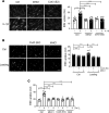
Figure 1. MHC1 antibody binds Cx43 and inhibits the opening of Cx43 hemichannels.
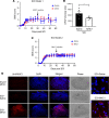
(A and B) Fixed parental HeLa cells or HeLa cells stably transfected with Cx43 were incubated with MHC1 antibody and then labeled with HRP-conjugated anti-human IgG (A) or rhodamine-conjugated anti-human IgG and counterstained with DAPI (B). Scale bars: 50 μm (A), 30 μm (B). (C) The binding affinity of MHC1 and IgG control to Cx43 peptide was determined by ELISA. n = 4. (D) Kinetics of MHC1 binding to the Cx43 extracellular domain peptide (N′-CFLSRPTEKTI) as assessed using an Octet RED96. (E) Parental HeLa cells or HeLa cells stably transfected with Cx43 were incubated with EGTA to remove extracellular Ca2+ ([Ca2+ ]0) in the absence or presence of MHC1 (66.7 nM) or control IgG (66.7 nM) before dye uptake assay with 15-minute treatment of 50 μM ethidium bromide (EtBr). (F) Primary astrocytes isolated from rat cortical brain were incubated for 3 or 24 hours with MHC1 (66.7 nM) or CBX (100 μM) before scrape-loading dye transfer assay was performed with Lucifer yellow (1%) and rhodamine dextran (1%) for 5 minutes. The level of EtBr dye uptake in E and dye transfer in F was determined by fluorescence microcopy and quantified by NIH ImageJ software. Scale bar: 200 μm (E and F). Data are presented as mean ± SEM of 3 independent experiments; each experiment had 2–3 repeats (2–3 wells). Michaelis-Menten equation was used in statistical analysis model (C). General linear model was used in statistical analysis (E and F). ***P < 0.001; ****P < 0.0001.
Figure 2
Figure 2. MHC1 antibody inhibits hemichannel opening in astrocytes by cytokine, mechanical stimulation, and low extracellular Ca2+/Mg2+.
(A) Primary astrocytes isolated from mouse spinal cord were treated with IL-1β (1 nM) for 24 hours. MHC1 (66.7 nM) or Cx43 (E2) antibody (6.7 nM) was then added 30 minutes prior to dye uptake assay with EtBr (50 μM) for 5 minutes. (B) Primary astrocytes isolated from mouse spinal cord were subjected to mechanical loading by Flexcell device in the absence or presence of MHC1 (66.7 nM) or Cx43E2 antibody (20 nM), and after loading, dye uptake assay was performed with EtBr (50 μM) for 5 minutes. (C) Primary astrocytes isolated from mouse spinal cord were incubated with or without EGTA to remove or maintain extracellular Ca2+/Mg2+ ([Ca2+ ]0) in the absence or presence of MHC1 (66.7 nM), Cx43E2 antibody (6.7 nM) or CBX (100 μM) for 30 minutes before dye uptake assay with EtBr (50 μM) for 15 minutes. Plus sign represents the presence of EGTA and absence of [Ca2+ ]0. Minus sign represents the absence of EGTA and presence of [Ca2+ ]0. The level of EtBr dye uptake was determined by fluorescence microcopy and quantified by NIH ImageJ software. The results are presented as mean ± SEM of 3 independent experiment, each experiment had 3–5 repeats (3–5 wells). Scale bar: 100 μm (A and B). General linear model was used in statistical analysis (AC). **P < 0.01, ****P < 0.0001.
Figure 3
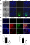
Figure 3. MHC1 delivered to spinal cord inhibits opening of hemichannels after acute SCI.
(A) MHC1 antibody (25 mg/kg) was i.p. injected 30 minutes after SCI under model 2. Four hours after the injection, spinal cords were isolated and fixed, and frozen tissue sections were prepared and immunolabeled with FITC-conjugated anti-human IgG secondary antibody. Images were taken from injury site. Scale bar: 100 μm. (B) MHC1 antibody (25 mg/kg) was i.p. injected 30 minutes after SCI under model 1. Evans blue (EB) and FITC-dextran dye were coinjected through tail vein 4 hours after i.p. injection. Mice were euthanized and perfused before isolation of spinal cords. Frozen tissue sections were prepared and EB dye uptake (red) was detected by fluorescence microscopy. Images were taken from the perilesional area (area < 1.5 mm from injury border). Scale bar: 50 μm. The percentage (C) and signal intensity (D) of EB-positive cells were quantified by NIH ImageJ software, and the results were combined from injury site, perilesional area, and distal area. The results are presented as mean ± SEM. SCI+Saline (n = 4), SCI+MHC1 (n = 4). Unpaired t test (1 tailed) was used in statistical analysis (C and D). **P < 0.01.
Figure 4
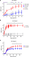
Figure 4. Mice with SCI recover hind limb function after treatment with Cx43 antibody.
After SCI, the hind limb function was evaluated with BMS, 1–9. 0 = no hind limb function and 9 = completely normal hind limb function. (A) Mice were subjected to broad impactor of SCI (model 1), and BMS score was measured for 12 days. MHC1 antibody–treated SCI mice were compared with IgG-treated SCI mice. Sham+Saline (n = 5), Sham+MHC1 (n = 5), SCI+Saline (n = 5), SCI+IgG (n = 8), SCI+MHC1 (n = 10). (B) Mice were subjected to broad impactor of SCI (model 1), and BMS score was measured for 56 days. SCI+IgG (n = 3), SCI+MHC1 (n = 5). (C) Mice were subjected to focused impactor of SCI (model 2), and BMS score was measured for 56 days. SCI+Saline (n = 15), SCI+MHC1 (n = 15). The data are presented as mean ± SEM. Statistical analysis consisted of linear mixed model of repeated measures, with antedependent covariance structure of time in days as best fit model (A); linear mixed model of repeated measures, with autoregressive covariance structure of time as best fit model (B); and 2-way repeated measures ANOVA with time as best fit model (C). *P < 0.05, **P < 0.01.
Figure 5
Figure 5. Blocking Cx43 hemichannel function by MHC1 antibody decreases spinal cord gliosis and protects neuron survival under the model 1 impact.
Spinal cords were isolated 14 days or 56 days after the model 1 impact of SCI treated with IgG control or MHC1 antibody. (A) The frozen sections were immunolabeled with anti-MAP2 or anti-GFAP antibody and counterstained with DAPI. The representative images of MAP2 and GFAP immunofluorescence were shown from the perilesion areas. (B) The quantification of MAP2-positive signals by NIH ImageJ software. SCI+IgG 14D (n = 3), SCI+MHC1 14D (n = 3), SCI+IgG 56D (n = 3), SCI+MHC1 56D (n = 3). (C) The quantification of GFAP-positive signals by NIH ImageJ software. SCI+IgG 14D (n = 3), SCI+MHC1 14D (n = 3), SCI+IgG 56D (n = 3), SCI+MHC1 56D (n = 4). All images were taken from perilesional area, which is confined to 1.5 mm from injury border of sagittal sections. The white dotted lines label the lesion border. Scale bar: 50 μm. Data are presented as mean ± SEM. Unpaired t test (1 tailed) was used in statistical analysis. **P < 0.01.
Figure 6
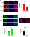
Figure 6. Blocking Cx43 hemichannel function by MHC1 antibody decreases spinal cord gliosis, protects neuron survival and reduces SCI lesion under the model 2 impact.
Spinal cords of SCI treated with saline control or MHC1 antibody were isolated 56 days after the model 2 impact. (A) The frozen sections were immunolabeled with anti-GFAP antibody and counterstained with DAPI. Left panels show representative images of GFAP immunofluorescence. Right panel show the quantification of GFAP-positive signals from perilesional area (area < 0.5 mm from injury border on sagittal sections) by NIH ImageJ software. Images were taken from perilesional area. White line labels the injury site border, and the area under the white lines is the lesion site. Scale bar: 0.5 mm. (B) The frozen sections were immunolabeled with anti-MAP2 antibody and counterstained with DAPI. Upper panels show representative images of MAP2 immunofluorescence, and lower panel shows the quantification of MAP2-positive signals from perilesional area (area < 0.5 mm from injury border on sagittal sections) by NIH ImageJ software. All images were taken from perilesional area. White line labels the injury site border, and the area under the white lines is the lesion site. Scale bar: 0.5 mm. (C) The white lines define SCI lesion areas (upper panel), and the sizes of lesion areas were quantified by NIH ImageJ software (lower panel). Scale bar: 50 μm. Data are presented as mean ± SEM. Unpaired t test (1 tailed) was used in statistical analysis. (AC). For GFAP and MAP2 staining, SCI+Saline (n = 6), SCI+MHC1 (n = 6). For lesion area, SCI+Saline (n = 5), SCI+MHC1 (n = 4). *P < 0.05, **P < 0.01, ***P < 0.001.
Figure 7
Figure 7. Blocking Cx43 hemichannel function by MHC1 antibody reduces SCI lesion and increases white matter and gray matter sparing under the model 2 impact.
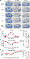
Spinal cords were isolated 30 days after the model 2 impact of SCI with the treatment of saline control or MHC1 antibody. The 10 μm thickness cryo-tissue sections were collected every 100 μm for a total 2 mm length centered on injury site, stained with Eriochrome cyanine R (A), and analyzed for lesion (B), white matter sparing (C), and gray matter sparing (D) by FIJI-ImageJ. The black dotted lines label the lesion border. Area under the curve was calculated. SCI+Saline (n = 10), SCI+MHC1 (n = 13). Scale bar: 500 μm. Data are presented as mean ± SEM. Unpaired t test (1 tailed) was used in statistical analysis. *P < 0.05.
Figure 8
Figure 8. Mice do not show improved recovery with Cx43 antibody 1 month after SCI.
Thirty days after SCI, mice were injected with 25 mg/kg MHC1 via intracerebroventricular route. (A) Mice were subjected to broad impact (model 1). BMS test was performed 30 days after MHC1 administration. SCI+Saline (n = 14), SCI+MHC1 (n = 16). (B) Mice were subjected to a broad impact (model 1). The frozen sections of spinal cords 60 days after SCI were immunolabeled with anti-GFAP antibody, and GFAP-positive signals from perilesional area (area < 0.5 mm from injury border on sagittal sections) were quantified using NIH ImageJ software. Data are presented as mean ± SEM. SCI+Saline (n = 5), and SCI+MHC1 (n = 5). (C) Mice were subjected to focused impact (model 2), and BMS score was determined in mice treated with MHC1 or saline control. SCI+Saline (n = 9), SCI+MHC1 (n = 10). (D) Mice were subjected to focused impact (model 2). Four hours after ICV injection with saline or MHC1, spinal cords were isolated; the frozen sections of spinal cords around perilesional sites were immunolabeled with anti-human IgG secondary antibody and counterstained with DAPI. Scale bar, 500 μm (black), 10 μm (white, right panels). Two-way repeated measures ANOVA with time was used for BMS statistical analysis (A and C). Unpaired t test (1 tailed) was used in statistical analysis (B). *P < 0.05.

Similar articles

Cited by

References

    1. Soden RJ, et al. Causes of death after spinal cord injury. Spinal Cord. 2000;38(10):604–610. doi: 10.1038/sj.sc.3101080. - DOI - PubMed
    1. Tator CH, Fehlings MG. Review of the secondary injury theory of acute spinal cord trauma with emphasis on vascular mechanisms. J Neurosurg. 1991;75(1):15–26. doi: 10.3171/jns.1991.75.1.0015. - DOI - PubMed
    1. Goldshmit Y, et al. Blood glutamate scavenger as a novel neuroprotective treatment in spinal cord injury. J Neurotrauma. 2018;35(21):2581–2590. doi: 10.1089/neu.2017.5524. - DOI - PubMed
    1. O’Carroll SJ, et al. Connexin 43 mimetic peptides reduce swelling, astrogliosis, and neuronal cell death after spinal cord injury. Cell Commun Adhes. 2008;15(1):27–42. - PubMed
    1. Hayashi M, et al. Sequential mRNA expression for immediate early genes, cytokines, and neurotrophins in spinal cord injury. J Neurotrauma. 2000;17(3):203–218. doi: 10.1089/neu.2000.17.203. - DOI - PubMed

MeSH terms