. 2021 Mar 8;6(5):e135633.
doi: 10.1172/jci.insight.135633.

α-Synuclein antisense oligonucleotides as a disease-modifying therapy for Parkinson's disease

Affiliations

α-Synuclein antisense oligonucleotides as a disease-modifying therapy for Parkinson's disease

Tracy A Cole et al. JCI Insight. .

Abstract

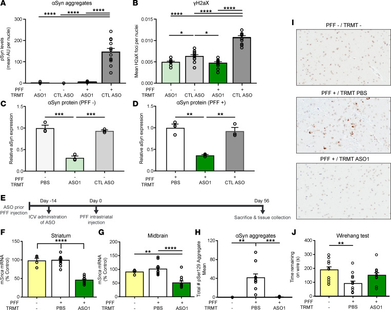
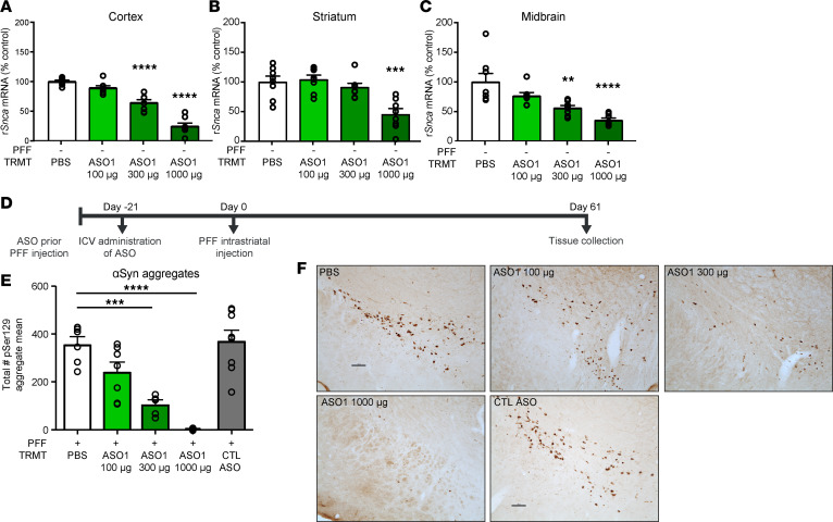
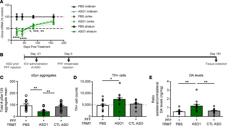
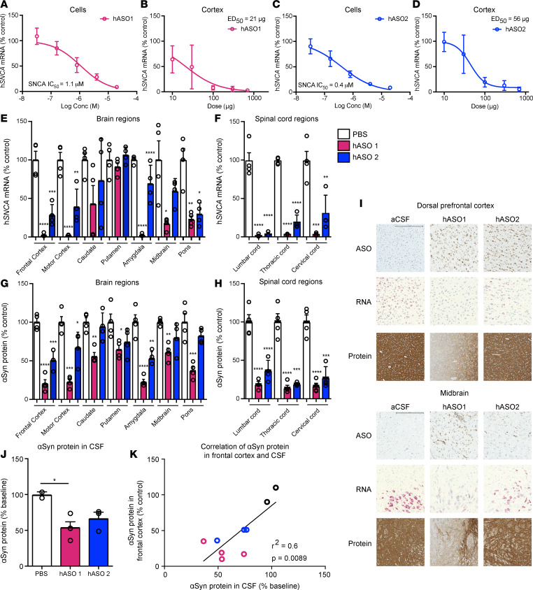
Parkinson's disease (PD) is a prevalent neurodegenerative disease with no approved disease-modifying therapies. Multiplications, mutations, and single nucleotide polymorphisms in the SNCA gene, encoding α-synuclein (aSyn) protein, either cause or increase risk for PD. Intracellular accumulations of aSyn are pathological hallmarks of PD. Taken together, reduction of aSyn production may provide a disease-modifying therapy for PD. We show that antisense oligonucleotides (ASOs) reduce production of aSyn in rodent preformed fibril (PFF) models of PD. Reduced aSyn production leads to prevention and removal of established aSyn pathology and prevents dopaminergic cell dysfunction. In addition, we address the translational potential of the approach through characterization of human SNCA-targeting ASOs that efficiently suppress the human SNCA transcript in vivo. We demonstrate broad activity and distribution of the human SNCA ASOs throughout the nonhuman primate brain and a corresponding decrease in aSyn cerebral spinal fluid (CSF) levels. Taken together, these data suggest that, by inhibiting production of aSyn, it may be possible to reverse established pathology; thus, these data support the development of SNCA ASOs as a potential disease-modifying therapy for PD and related synucleinopathies.

Keywords: Neuroscience; Parkinson disease; Therapeutics.


Conflict of interest statement

Conflict of interest: TAC, HZ, AM, CFB, DQ, EES, and HBK are paid employees and stockholders of Ionis Pharmaceuticals Inc. DG, MW, MB, LJ, and AW are paid employees and stockholders of Biogen.

Figures

Figure 1
Figure 1. ASO-mediated reduction of Snca improves cellular function in cells and prevents pathogenic aSyn aggregate deposition in an in vivo PFF model of PD.
(A and B) Quantification of pSer129+ area by intensity in mouse primary cortical cultures and cellular function, by γH2AX Ser139, in mouse primary cortical cultures with either CTL ASO or ASO1 30 minutes following PFF addition. Replicated 2 times. (C) Quantification of aSyn protein by Western blot from mouse primary cortical cultures not treated with PFF (n = 3). (D) Quantification of aSyn protein by Western blot from mouse primary cortical cultures treated with PFF. (E) Timeline for single 700 μg i.c.v. bolus ASO administration prior to the PFF injection paradigm with termination at day 56. (F and G) mRNA reduction by RT-PCR in striatum and midbrain. (H) Quantification of pSyn+ aggregate reduction (total enumeration) in the substantia nigra by IHC. (I) Representative images of immunostaining (IHC) for pSer129+ aggregate counts. Original magnification, 100×. (J) Performance on a wire hang task (n = 4, 12, and 11 for naive, PBS, and ASO1, respectively, except wire hang, in which n = 12 for naive). Data are represented as ± SEM. *P < 0.05, **P < 0.01, ***P < 0.001, ****P < 0.0001 (2-way ANOVA with Tukey post hoc analyses for duration of action with all other analyses using 1-way ANOVA with Tukey post hoc analyses). PFF, preformed fibril; TRMT, treatment.
Figure 2
Figure 2. ASO-mediated reduction of Snca is dose responsive and prevents pathogenic aSyn aggregate deposition in an in vivo PFF model of PD.
(AC) Three-week dose response of rat Snca levels from cortical, striatal, and midbrain rat samples by qPCR (n = 8 per dose). (D) Timeline for ASO dose-response administration prior to PFF injection paradigm. (E) Quantification of immunostaining for pSer129+ aggregate counts by total enumeration (n = 6, 7, 5, 8, 8 for PBS, 100 μg, 300 μg, 1000 μg, and CTL ASO, respectively). (F) Representative images of pSer129+ aggregate counts in the substantia nigra. Scale bar: 100 μm. Data are represented as ± SEM. **P < 0.01, ***P < 0.001, ****P < 0.0001 (2-way ANOVA with Tukey post hoc analyses for duration of action, with all other analyses using 1-way ANOVA with Tukey post hoc analyses). PFF, preformed fibril; TRMT, treatment; CTL ASO, control ASO.
Figure 3
Figure 3. ASO-mediated suppression of Snca exhibits a prolonged duration of action and prevents dopaminergic cell dysfunction in an in vivo PFF model of PD.
(A) Time course of Snca mRNA reduction (the 1000 μg results for the 3-week time point in Figure 2, A–C are included). (BE) Results from ASO administration (1000 μg) prior to PFF injection paradigm in rats with study termination at 181 days. (B) Timeline for ASO administration prior to PFF injection paradigm in rats. (CE) pSer129+ aggregate counts using total enumeration by IHC (n = 13, 13, and 15 for PBS, ASO1, and CTL ASO, respectively), dopaminergic cell counts by IHC (by stereology) (n = 13, 13, and 12 for PBS, ASO1, and CTL ASO, respectively), and striatal dopamine levels by HPLC normalized to the contralateral side (n = 13, 13, and 14 for PBS, ASO1, and CTL ASO, respectively). Data are represented as ± SEM. *P < 0.05, **P < 0.001, ***P < 0.0001, ****P < 0.00001 (1-way ANOVA with Tukey post hoc analyses). PFF, preformed fibril; TRMT, treatment; CTL ASO, control ASO. Statistical significance was also achieved when using nonparametric test Kruskall-Wallis for Figure 3, D and E; P ≥ 0.02. The same animal was high for TH and DA levels.
Figure 4
Figure 4. Pathogenic aSyn aggregate deposition is reversible, and its amelioration prevents TH loss.
(A) Timeline for ASO administration after PFF injection paradigm in the mouse. (B and C) mRNA reduction by RT-PCR (n = 12, 10, and 10 for naive, PBS, and ASO1, respectively) (D and E) Quantification of aggregate reduction in the substantia nigra by IHC (n = 4, 10, and 10 for naive, PBS, and ASO1, respectively) and performance on a wire hang task (n = 10, 10, and 10 for naive, PBS, ASO1, respectively). (F) Timeline for ASO administration (1000 μg) for GI. (G) Quantification of Snca mRNA reduction in the midbrain at each time point. (H and I) Quantification of immunostaining for pSer129+ aggregate counts in the midbrain, and representative images from the insular cortex (n = 10, 9, 9, 9, and 10 for PFF only, PBS 60 days, ASO1 60 days, PBS 81 days, and ASO1 81 days, respectively). (JM) Results from ASO administration (1000 μg) with study termination at 181 days. (J) Timeline for ASO administration for KM. (K) TH+ cell counts by IHC (by stereology) (n = 12, 11, and 14 for PBS, ASO1, and CTL ASO, respectively). (L and M) Quantification of pSer129+ aggregate counts in the substantia nigra by IHC (n = 13, 12, and 12 for PBS, ASO1, and CTL ASO, respectively) and striatal dopamine levels by HPLC normalized to the contralateral side (n = 13, 12, and 14 for PBS, ASO1, and CTL ASO, respectively). Data are represented as ± SEM. *P < 0.05, **P < 0.001, ***P < 0.0001, ****P < 0.00001 (1-way ANOVA with Tukey post hoc analyses). PFF, preformed fibril; TRMT, treatment; CTL ASO, control ASO.
Figure 5
Figure 5. Sustained reduction of Snca with ASO administered prior to and after establishment of aggregates reduces aggregate pathology and prevents TH loss.
(AF) Results from ASO administration prior to PFF injection paradigm with sustained suppression and study termination at 224 days after PFF injection in mice. (A) Timeline for ASO administration (700 μg) prior to PFF injection paradigm. (B and C) mRNA reduction in the striatum and midbrain. (D and E) pSer129+ aggregate counts quantified by IHC in neurites and cell bodies. (F) Dopaminergic cell counts quantified by IHC. (n = 12 for PBS and ASO1; n = 6 for IHC endpoints). (GK) Results from ASO administration after PFF injection paradigm with sustained suppression and study termination at 224 days after PFF injection in mice (700 μg). (G) Timeline for ASO administration (700 μg) after PFF injection paradigm. (H and I) mRNA reduction in the striatum and midbrain (n = 4, 6, 4, respectively, for PBS, ASO1, and CTL ASO). (J) pSer129+ aggregate counts quantified by IHC (n = 4). (K) Dopaminergic cell counts quantified by IHC. (L) Representative images of aSyn IHC from coronal sections (n = 4). Scale bar: 20 µm. Data are represented as ± SEM. *P < 0.05, **P < 0.001, ***P < 0.0001, ****P < 0.00001 (1-way ANOVA with Tukey post hoc analyses). PFF, preformed fibril; TRMT, treatment; CTL ASO, Control ASO.
Figure 6
Figure 6. Human SNCA ASOs are potent, suppress aSyn broadly in the primate CNS, and CSF aSyn is a suitable pharmacodynamic biomarker.
(AD) In vitro dose response of human SNCA hASO1 and hASO2 in SHSY5Y cells and in vivo dose response in SNCA-PAC mouse cortex 2 weeks after injection (n = 10, except 700 μg n = 7 for hASO1; n = 8 for hASO2 [2 mice removed with ROUT analysis from 300 μg and 700 μg groups], combination of 2 studies of n = 4 each; n = 12 for all PBS groups). (EH) mRNA by RT-PCR and protein by ELISA throughout the brain and spinal cord of the NHP (n = 4 for PBS, hASO1, and hASO2). (I) Representative images for IHC (ASO and aSyn antibody) or ISH (SNCA mRNA). Scale bar: 600 μm for protein/300 μm for ASO and ISH results. (J and K) Quantification of aSyn protein by ELISA in CSF with correlation of aSyn protein levels in the frontal cortex and CSF (n = 2, 3, and 4 for PBS, hASO1, and hASO2, respectively). Data are represented as ± SEM, except for SHSY5Y, which are presented as mean ± SD. *P < 0.05, **P < 0.001, ***P < 0.0001, ****P < 0.00001 (1-way ANOVA with Tukey post hoc analyses).

References

    1. Chartier-Harlin MC, et al. Alpha-synuclein locus duplication as a cause of familial Parkinson’s disease. Lancet. 2004;364(9440):1167–1169. doi: 10.1016/S0140-6736(04)17103-1. - DOI - PubMed
    1. Devine MJ, et al. Parkinson’s disease and α-synuclein expression. Mov Disord. 2011;26(12):2160–2168. doi: 10.1002/mds.23948. - DOI - PMC - PubMed
    1. Farrer M, et al. Comparison of kindreds with parkinsonism and alpha-synuclein genomic multiplications. Ann Neurol. 2004;55(2):174–179. doi: 10.1002/ana.10846. - DOI - PubMed
    1. Fuchs J, et al. Phenotypic variation in a large Swedish pedigree due to SNCA duplication and triplication. Neurology. 2007;68(12):916–922. doi: 10.1212/01.wnl.0000254458.17630.c5. - DOI - PubMed
    1. Fuchs J, et al. Genetic variability in the SNCA gene influences alpha-synuclein levels in the blood and brain. FASEB J. 2008;22(5):1327–1334. doi: 10.1096/fj.07-9348com. - DOI - PubMed

MeSH terms