

doi: 10.3791/62183.
Creating Avian Forebrain Chimeras to assess Facial Development
Affiliations
- PMID: 33682859
- PMCID: PMC8628319
- DOI: 10.3791/62183
Item in Clipboard
Creating Avian Forebrain Chimeras to assess Facial Development
J Vis Exp.
.
Abstract
The avian embryo has been used as a model system for more than a century and has led to fundamental understanding of vertebrate development. One of the strengths of this model system is that the effect of, and interaction among, tissues can be directly assessed in chimeric embryos. We have previously shown that signals from the forebrain contribute to facial morphogenesis by regulating the shape of the expression domain of Sonic hedgehog (SHH) in the Frontonasal Ectodermal Zone (FEZ). In this article, the method of generating the forebrain chimeras and provide illustrations of the outcomes of these experiments is described.
Figures
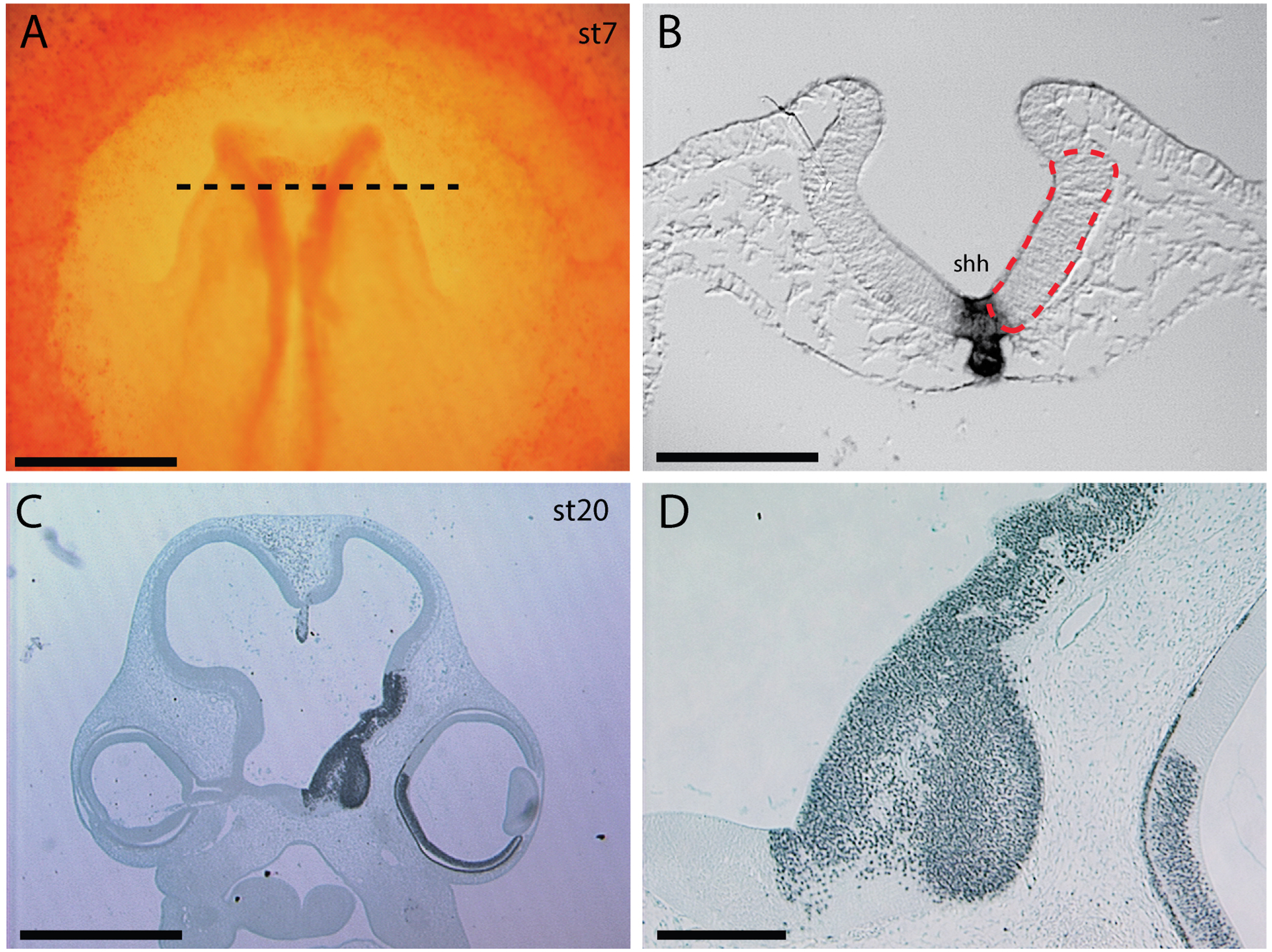
(A) Dorsal view of stage 8 chicken embryo stained with neutral red showing the location of the graft and engraftment site. Dotted line is approximate level shown in B. (B) Cross section through a stage 8 chicken embryo after in situ hybridization to show SHH expression. The approximate location of the graft is shown, red dotted box. (C) Immunostaining to detect QCPN in quail-chick chimeras shows the graft is widespread and contributes only to the ventro-lateral neural tube (arrow) and ventral optic cup (arrowhead). (D) Higher magnification in C. Scale bars: A: 500um, B: 100um, C: 1mm, D: 200um.

(A, B) Two examples of quail-duck chimeras at stage 22. These embryos have serious malformations that preclude further analysis. Scale bar: 2mm

(A) Normal chick and (B) duck embryos at stage 22 after whole mount in situ hybridization to visualize SHH expression. (C) A chick-chick control shows a pattern of SHH and morphology that resembles a normal chick embryo. (D) A duck-chick chimera shows an altered pattern of SHH expression. On the transplanted side SHH expression (yellow dotted line) is more rounded and resembles the duck pattern. The nasal pit is also more rounded on the transplanted side (yellow arrow) compared to the more “slit” like appearance on the host side (red arrow). The red bar indicates the midline and the transplant is on the right side of the image. Scale bar: 1mm
References
-
- Waddington C Developmental Mechanics of Chicken and Duck Embryos.. Nature 125, 924–925 (1930).
Publication types
MeSH terms
Grants and funding
LinkOut - more resources
Full Text Sources
Other Literature Sources
