. 2021 Jun 4;104(6):1337-1346.
doi: 10.1093/biolre/ioab036.

Neurotensin: a neuropeptide induced by hCG in the human and rat ovary during the periovulatory period†

Affiliations

Neurotensin: a neuropeptide induced by hCG in the human and rat ovary during the periovulatory period†

Linah Al-Alem et al. Biol Reprod. .

Abstract

Neurotensin (NTS) is a tridecapeptide that was first characterized as a neurotransmitter in neuronal cells. The present study examined ovarian NTS expression across the periovulatory period in the human and the rat. Women were recruited into this study and monitored by transvaginal ultrasound. The dominant follicle was surgically excised prior to the luteinizing hormone (LH) surge (preovulatory phase) or women were given 250 μg human chorionic gonadotropin (hCG) and dominant follicles collected 12-18 h after hCG (early ovulatory), 18-34 h (late ovulatory), and 44-70 h (postovulatory). NTS mRNA was massively induced during the early and late ovulatory stage in granulosa cells (GCs) (15 000 fold) and theca cells (700 fold). In the rat, hCG also induced Nts mRNA expression in intact ovaries and isolated GCs. In cultured granulosa-luteal cells (GLCs) from IVF patients, NTS expression was induced 6 h after hCG treatment, whereas in cultured rat GCs, NTS increased 4 h after hCG treatment. Cells treated with hCG signaling pathway inhibitors revealed that NTS expression is partially regulated in the human and rat GC by the epidermal-like growth factor pathway. Human GLC, and rat GCs also showed that Nts was regulated by the protein kinase A (PKA) pathway along with input from the phosphotidylinositol 3- kinase (PI3K) and mitogen-activated protein kinase (MAPK) pathways. The predominat NTS receptor present in human and rat GCs was SORT1, whereas NTSR1 and NTSR2 expression was very low. Based on NTS actions in other systems, we speculate that NTS may regulate crucial aspects of ovulation such as vascular permeability, inflammation, and cell migration.

Keywords: cumulus oocyte complex; fertility; granulosa cell; neurotensin; ovary; ovulation.


Figures

Figure 1
Figure 1
Neurotensin (NTS) mRNA expression across the ovulatory period in the human. Data are presented as the fold change relative to the preovulatory (Pre, no hCG) time point. NTS mRNA increases in the early and late ovulatory phase in the human granulosa (A) and theca (B) cells. Results are the means ± standard error of mean of n = 4–5. A one-way analysis of variance was used to test differences across the time points for the granulosa and theca cells. The Tukey post hoc test was performed and bars that do not share a letter designation are significantly different (p < 0.05). Immunostaining of NTS protein expression across the different stages of the human ovulatory cycle demonstrates low expression of NTS prior to hCG (C). Expression is present in the granulosa cells (gc) during the early (D) and late ovulatory periods (E). an = antrum, st = stroma.
Figure 2
Figure 2
Neurotensin (NTS) mRNA expression in cumulus cells of matched GLC patients. NTS expression is higher in cumulus cells when normalized to GLC from matched patients collected at the time of oocyte retrieval as analyzed by the Student t-test (A). When taken collectively, there is a > 200 fold increased NTS expression compared with GLC at the time of collection (B).
Figure 3
Figure 3
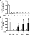
Neurotensin (NTS) mRNA expression in human GLC in vitro. Human GLCs were collected at the day of IVF retrieval (day 0) and NTS expression was examined across the 7 days of culture (A). After 6–7 days of culture, GLCs were serum starved for 1 h and then treated with vehicle control (−) or hCG (+) for 24 h (B). “Coll” represents samples at the time of IVF collection. A total of 0 h represents samples after 6–7 days in culture at the initiation of hCG treatment. Results are the means ± standard error of mean of n = 3. A two-way analysis of variance was used to test differences with or without hCG treatment across the time points. The Tukey post hoc test was performed and bars that do not share a letter designation are significantly different (p < 0.05).
Figure 4
Figure 4
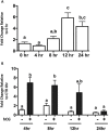
Nts mRNA expression across the ovulatory period in the rat. Nts expression in intact ovaries increases during the late ovulatory period prior to follicular rupture (12 h) and remains elevated (A). Nts mRNA expression in isolated rat granulosa cells in vitro is stimulated by 4 h after hCG and declines to control levels by 24 h (A). Results are the means ± standard error of mean of n = 4. A one-way analysis of variance (ANOVA) was used to test differences across the time points in intact ovaries. A two-way ANOVA was used to test differences across the time points with or without hCG treatment in isolated granulosa cells. The Tukey post hoc test was performed and bars that do not share a letter designation are significantly different (p < 0.05).
Figure 5
Figure 5
Regulation of Neurotensin (NTS) mRNA expression in human GLC and rat granulosa cells in vitro. Inhibitors for hCG signaling pathways were used to determine which signaling pathway is involved in the expression of NTS. Rat granulosa cells (A) and human granulosa-luteal cells (B) were incubated for 1 h with the following hCG pathway inhibitors: AG (AG1478; EGF receptor inhibitor), RU (RU486; progesterone receptor antagonist), NS (NS398; prostaglandin synthase 2 inhibitor) or IND (Indomethacin; a prostaglandin synthase 1 and 2 inhibitor). The hCG induction of NTS mRNA expression was partially by the EGF receptor inhibitor. Rat granulosa cells (C) and human GLC (D) were incubated for 1 h with the following signaling inhibitors: GF (GF109203x; PKC inhibitor), LY (LY294002; PI3K inhibitor), H89 (PKA inhibitor), SB (SB203580; p38 MAPK inhibitor) or U126 (MEK1/2 inhibitor). The hCG induction of NTS mRNA expression was regulated by the PKA pathway in both species. However differences existed in other signaling pathways between the species. Results are the means ± standard error of mean of n = 6. A two-way analysis of variance was used to test differences with or without hCG treatment across the different signaling pathway inhibitors. The Tukey post hoc test was performed and bars that do not share a letter designation are significantly different (p < 0.05).
Figure 6
Figure 6
Expression of Neurotensin (NTS) receptors in human and rat granulosa cells in vitro. The expression of mRNA after treatment with hCG is shown for NTSR2 (A) and SORT1 (B) in human granulosa collected across the periovulatory period. NTSR2 (C) and SORT1 (D) mRNA expression is also shown in cultured human GLCs. Human GLCs were cultured for 7 days and then treated with or without hCG. The expression of NTS receptors was also determined in isolated rat GC treated with or without hCG. The expression of mRNA for Ntsr2 (E) and Sort1 (F) after temporal treatment with hCG is shown. Results are the means ± standard error of mean of n = 3–5. A two-way analysis of variance was used to test differences with or without hCG treatment across the time points. The Tukey post hoc test was performed and bars that do not share a letter designation are significantly different (p < 0.05).

References

    1. Leeman SE, Carraway RE. Neurotensin: discovery, isolation, characterization, synthesis and possible physiological roles. Ann N Y Acad Sci 1982; 400:1–16. - PubMed
    1. Dungan Lemko HM, Naderi R, Adjan V, Jennes LH, Navarro VM, Clifton DK, Steiner RA. Interactions between neurotensin and GnRH neurons in the positive feedback control of GnRH/LH secretion in the mouse. Am J Physiol Endocrinol Metab 2010; 298:E80–E88. - PMC - PubMed
    1. Enjalbert A, Arancibia S, Priam M, Bluet-Pajot MT, Kordon C. Neurotensin stimulation of prolactin secretion in vitro. Neuroendocrinology 1982; 34:95–98. - PubMed
    1. Leeman SE. Substance P and neurotensin: discovery, isolation, chemical characterization and physiological studies. J Exp Biol 1980; 89:193–200. - PubMed
    1. Boules MM, Fredrickson P, Muehlmann AM, Richelson E. Elucidating the role of neurotensin in the pathophysiology and management of major mental disorders. Behav Sci (Basel) 2014; 4:125–153. - PMC - PubMed

Publication types