

Long non-coding RNA KCNQ1OT1 promotes the progression of gastric cancer via the miR-145-5p/ARF6 axis
- PMID: 33682985
- PMCID: PMC8244094
- DOI: 10.1002/jgm.3330
Long non-coding RNA KCNQ1OT1 promotes the progression of gastric cancer via the miR-145-5p/ARF6 axis
Retraction in
-
RETRACTION: Long non-coding RNA KCNQ1OT1 Promotes the Progression of Gastric Cancer via the miR-145-5p/ARF6 Axis.J Gene Med. 2024 Aug;26(8):e3727. doi: 10.1002/jgm.3727. J Gene Med. 2024. PMID: 39115084 Free PMC article. No abstract available.
Abstract
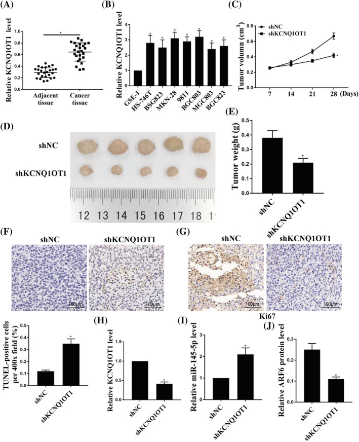
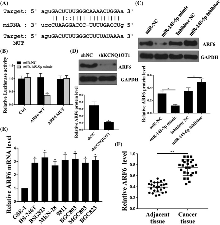
Background: Long non-coding RNA KCNQ1 opposite strand/antisense transcript one gene (KCNQ1OT1) has been reported to be involved in the progression of many types of human cancer, whereas its role in gastric cancer (GC) remains unknown. The present study aimed to investigate the role of KCNQ1OT1 in GC.
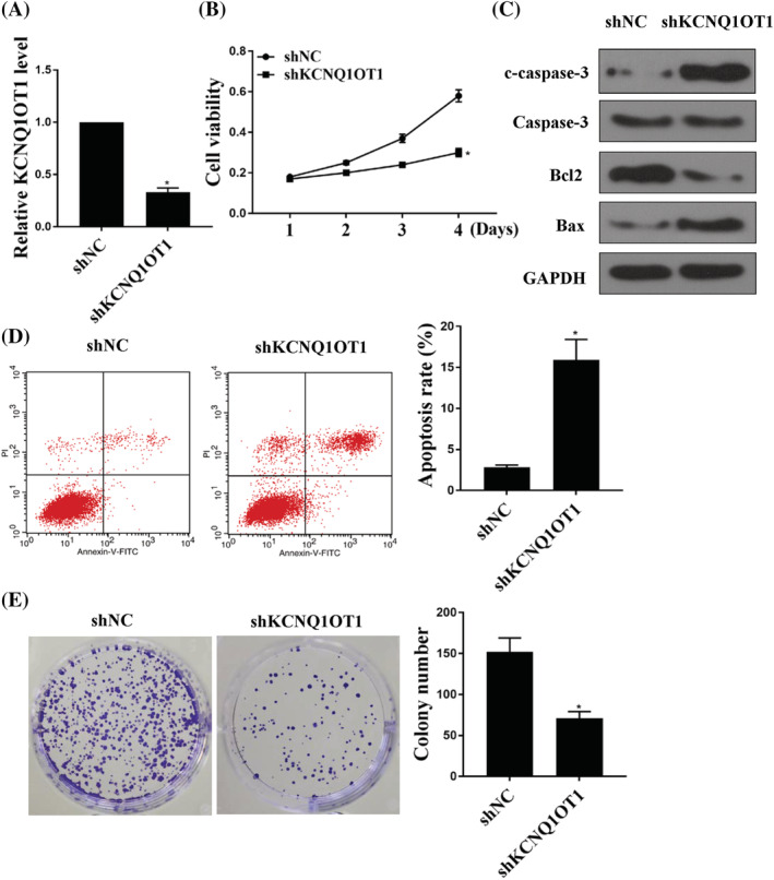
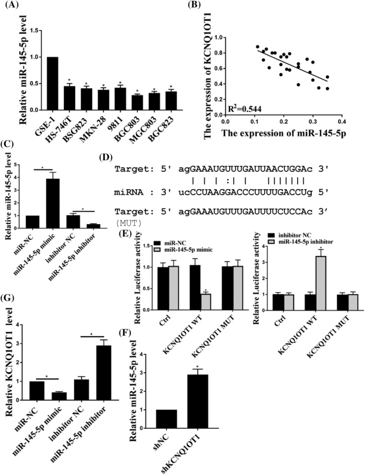
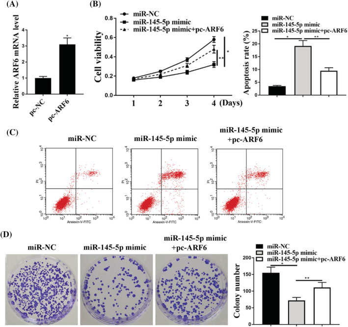
Methods: In total, 25 GC tissues and adjacent normal tissues were collected. The expression of KCNQ1OT1, miR-145-5p and ARF6 in GC tissues and cell lines was detected by quantitative reverse transcriptase-polymerase chain reaction or western blotting. Bioinformatics analysis and a dual luciferase reporter assay were performed to determine the relationship between KCNQ1OT1 and miR-145-5p or miR-145-5p and ARF6. Gain- and loss-of function of KCNQ1OT1 and miR-145-5p were achieved to confirm their roles in GC cells. Cell counting kit-8, colony formation and flow cytometry assays were used to evaluate cell viability, proliferation and apoptosis. A xenograft tumor model was established with BGC803 tumor cells transfected with sh-KCNQ1OT1 or empty vector to determine the role of LINC01089 in vivo.
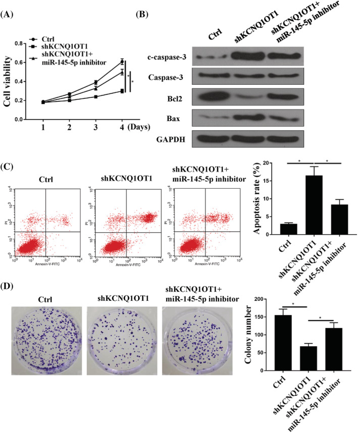
Results: The expression levels of KCNQ1OT1 were markedly elevated in GC tissues and cells. Knockdown of KCNQ1OT1 inhibited GC tumor growth, reduced GC cell viability and colony formation, and induced GC cell apoptosis. The expression levels of miR-145-5p were significantly decreased in GC cells and correlated with the expression of KCNQ1OT1 in GC tumors. Moreover, KCNQ1OT1 directly binds with miR-145-5p, which is targeting ARF6. Knockdown of KCNQ1OT1 increased the expression levels of miR-145-5p. Inhibition of miR-145-5p increased the expression levels of KCNQ1OT1 and also attenuated the effects of knockdown of KCNQ1OT1 on the viability, proliferation and apoptosis of GC cells. In addition, overexpression of miR-145-5p reduced GC cell viability and colony formation and induced GC cell apoptosis, whereas overexpression of ARF6 attenuated the effects of overexpression of miR-145-5p on GC cell viability, colony formation and apoptosis.
Conclusions: KCNQ1OT1 can promote GC progression through the miR-145-5p/ARF6 axis. KCNQ1OT1 may serve as a therapeutic target and a diagnostic biomarker of GC.
Keywords: ARF6; KCNQ1OT1; gastric cancer; miR-145-5p.
© 2021 The Authors. The Journal of Gene Medicine published by John Wiley & Sons Ltd.
Conflict of interest statement
The authors declare that they have no conflicts of interest.
Figures
References
-
- Sun M, Nie F, Wang Z, De W. Involvement of lncRNA dysregulation in gastric cancer. Histol Histopathol. 2016;31:33‐39. - PubMed
-
- Muro K, Van Cutsem E, Narita Y, et al. Pan‐Asian adapted ESMO Clinical Practice Guidelines for the management of patients with metastatic gastric cancer: a JSMO–ESMO initiative endorsed by CSCO, KSMO, MOS, SSO and TOS. Ann Oncol. 2018;30:19‐33. - PubMed
-
- Hwang B, Lee S, Jung C, Yu B, Lee Y. Regulation and functional roles of autophagy in Helicobacter pylori CagA‐mediated gastric cancer. In: Proceedings: AACR Annual Meeting 2019; March 29‐April 3, 2019; Atlanta, GA.
-
- Plummer M, Franceschi S, Vignat J, Forman D, de Martel C. Global burden of gastric cancer attributable to Helicobacter pylori. Int J Cancer. 2015;136:487‐490. - PubMed
-
- Chen W, Zheng R, Baade PD, et al. Cancer statistics in China, 2015. CA Cancer J Clin. 2016;66:115‐132. - PubMed
Publication types
MeSH terms
Substances
LinkOut - more resources
Full Text Sources
Other Literature Sources
Medical
Miscellaneous
