Review
. 2021 Mar 8;12(1):32.
doi: 10.1186/s13244-021-00978-8.

Osteoid osteoma: the great mimicker

Affiliations
Review

Osteoid osteoma: the great mimicker

Bruno C Carneiro et al. Insights Imaging. .

Abstract

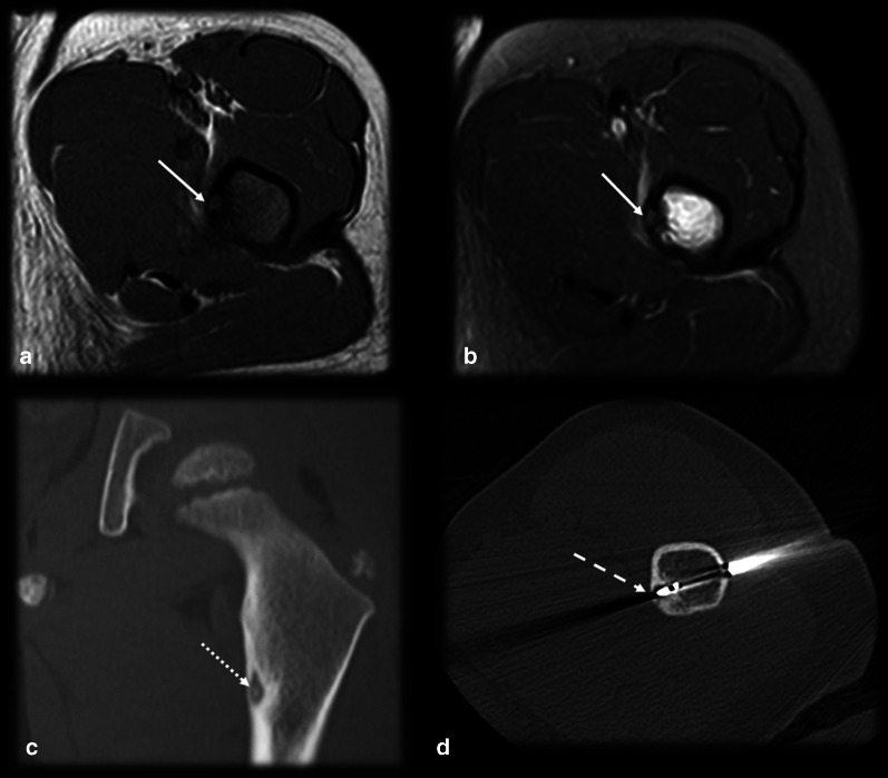
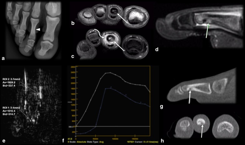
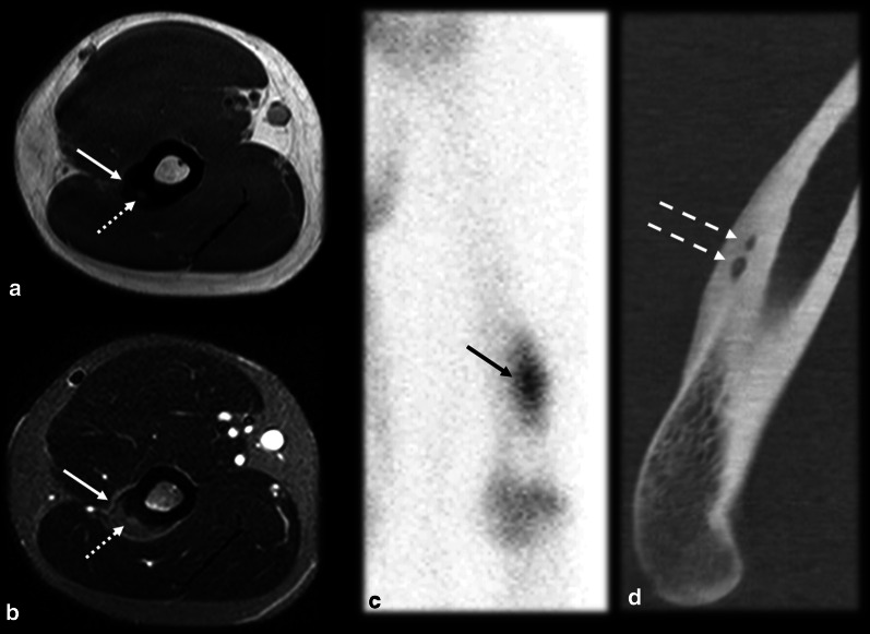
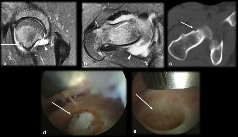
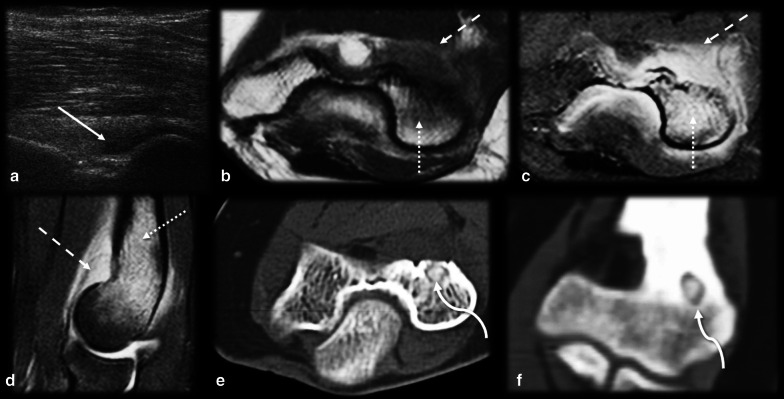
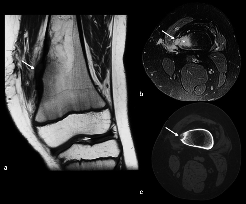
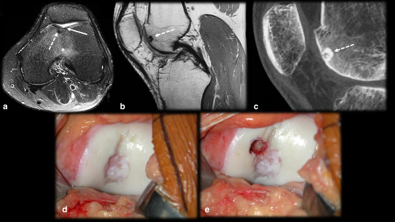
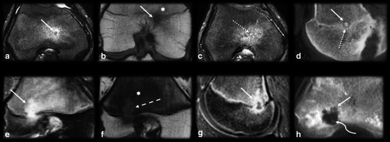
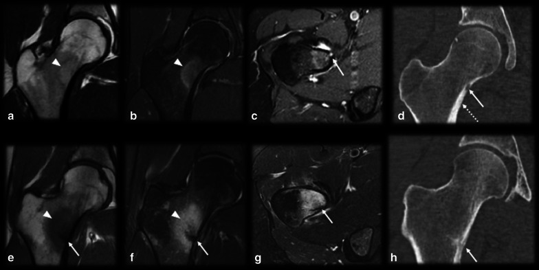
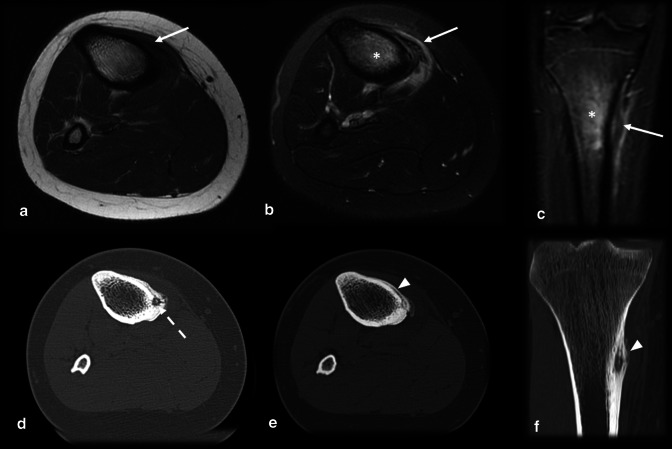
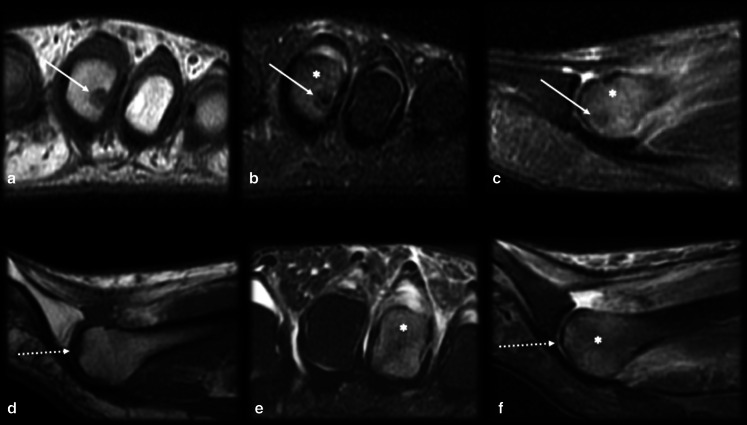
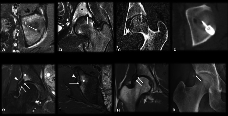
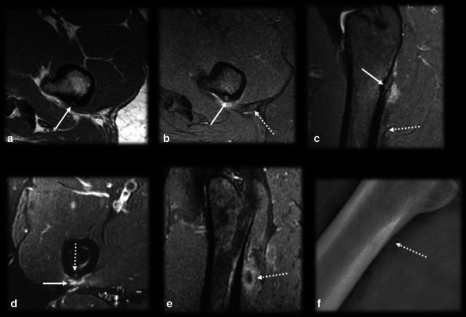
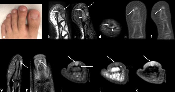
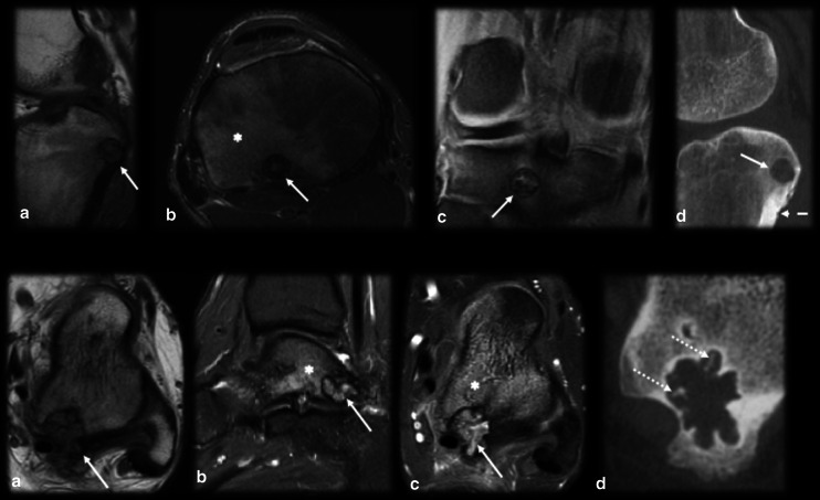
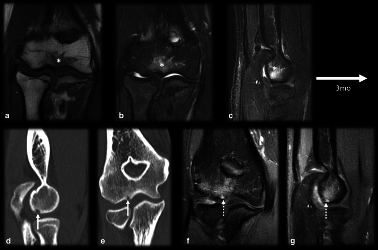
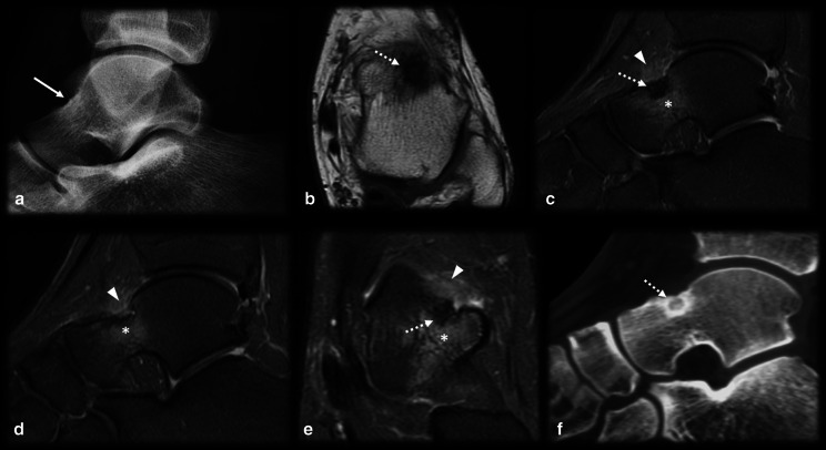
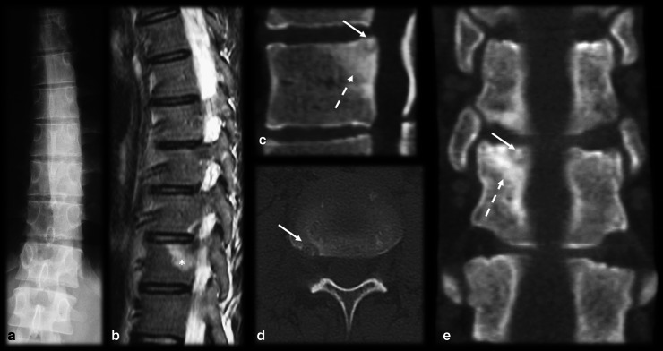
Osteoid osteoma is a painful, benign and common bone tumor that is prevalent in young adults. The typical clinical presentation consists of pain that becomes worse at night and is relieved by nonsteroidal anti-inflammatory drugs. The most common imaging finding is a lytic lesion, known as a nidus, with variable intralesional mineralization, accompanied by bone sclerosis, cortical thickening and surrounding bone marrow edema, as well as marked enhancement with intravenous contrast injection. When the lesion is located in typical locations (intracortical bone and the diaphyses of long bones), both characteristic clinical and radiological features are diagnostic. However, osteoid osteoma is a multifaceted pathology that can have unusual presentations, such as intraarticular osteoid osteoma, epiphyseal location, lesions at the extremities and multicentric nidi, and frequently present atypical clinical and radiological manifestations. In addition, many conditions may mimic osteoid osteoma and vice versa, leading to misdiagnosis. Therefore, it is essential to understand these musculoskeletal diseases and their imaging findings to increase diagnostic accuracy, enable early treatment and prevent poor prognosis.

Keywords: Bone neoplasms; Diagnosis; Differential; Magnetic resonance imaging; Osteoid; Osteoma; Tomography; X-ray computed.


Conflict of interest statement

The authors declare that they have no competing interests.

Figures

Fig. 1
Fig. 1
Typical osteoid osteoma and percutaneous ablation. Male, 15 years old, presenting with medial hip pain for 2 months, which became worse at night. Axial T1 (a) and T2 FS (b) MR images showing a small cortical nidus (arrows) within the femur shaft with a target-like appearance, edema and sclerosis. CT (c) better detected the partially mineralized nidus (dotted arrow), cortical thickening and sclerosis than did MRI. CT percutaneous biopsy and radiofrequency ablation (d) were performed (dashed arrow)
Fig. 2
Fig. 2
Pattern of enhancement of osteoid osteoma. Male, 17 years old, presenting with metatarsalgia of the right foot for 2 months, which became worse over the past week. He had no recollection of trauma and practiced sports regularly. CR (a) showed no significant findings, apart from mild bone sclerosis in the middle phalanx of the second toe (arrowhead). Coronal T1 and T2WI (b, c) depicted marked edema of the bone marrow and surrounding tissues (curved arrow) and a very small intracortical lesion (dashed arrow). Dynamic MR angiography (df) showed marked enhancement of the intracortical nodule (arrow in d), presenting contrast kinetics similar to those of adjacent arteries, with a peak enhancement followed by rapid washout, suggestive of OO (e, f). A CT scan (g, h) was later performed, and the findings confirmed the presence of a nidus (arrows)
Fig. 3
Fig. 3
Multicentric osteoid osteoma. A 26-year-old male handball player with lateral elbow and arm pain for 3 months. Orthopedists suspected lateral epicondylitis or a stress reaction. MRI T1 (a) and T2 FS (b) showed cortical thickening on the lateral supracondylar crest with corticoperiosteal edema (arrow) and small foci of intermediate signal intensity (dotted arrow), which raised the suspicion for OO. Scintigraphy (c) evidenced the double density sign (black arrow), which cannot be used to distinguish between single and multicentric OO. CT (d) detected two nidi (dashed arrows), confirming the diagnosis of multicentric OO
Fig. 4
Fig. 4
Osteoid osteoma mimicking synovitis of the hip. A 24-year-old man with right hip pain, swelling and tenderness for 2 weeks. Inflammatory marker levels were also elevated. Orthopedists suspected inflammatory arthropathy, and ultrasound (not shown) depicted joint effusion and synovitis. MRI T2 FS sagittal (a) and axial (b) shows joint effusion (arrowhead) with synovial thickening and a doubtful nidus (arrow), which was better characterized on the CT scan (c). Arthroscopic aspect before (d) and after (e) nidus resection
Fig. 5
Fig. 5
Osteoid osteoma mimicking synovitis on the elbow. Female, 20 years old, with elbow pain and edema for 4 months. Ultrasound in anterior sagittal view (a) at the coronoid fossa level shows joint effusion (arrow). MRI T1 (b) and T2FS (c, d) show bone marrow edema (dotted arrows) and synovitis (dashed arrows). Further investigation with a CT scan (e, f) revealed a mineralized nidus (curved arrow) in the cancellous bone of the medial humeral condyle
Fig. 6
Fig. 6
Osteoid osteoma near a growth plate. An 11–year-old male with a history of surgical removal of an OO on the distal metadiaphysis of the left femur. His symptoms persisted, and follow-up MRI (a, b) and CT (c) showed a residual nidus at the medial femoral margin represented by the intracortical nodule (arrows) and surrounding bone marrow edema (asterisk in b). In a, note that the affected side of the distal femur is longer than the lateral side, resulting in a femoral deformity and length discrepancy of the left lower limb
Fig. 7
Fig. 7
Subchondral osteoid osteoma mimicking trochlear chondromalacia. A 35-year-old male with anterior knee pain for 4 months. Axial T2 FS MR images (a) showed deep chondral erosion (arrow), subchondral edema (asterisk) and a small, low signal intensity foci that could be a nidus (dashed arrow). Sagittal T1 MRI (b) and CT (c) confirmed the diagnosis of the OO nidus (dashed arrow). Surgical images before (d) and after (e) resection
Fig. 8
Fig. 8
Osteoid osteoma versus osteomyelitis. (ad) Male, 15 years old, presenting with knee pain for 2 months. MRI T2 FS (a), T1 (b), T1 FSGD (c) and CT (d) showed a nidus with smooth margins (arrows), a central mineralized portion (dashed arrows), homogeneous gadolinium enhancement (dotted arrows) and hazy T1 bone marrow edema around the lesion (asterisk). (eh) Male, 13 years old, presenting with knee pain for 6 weeks. MRI T2 FS (e), T1 (f), T1 FS GD (g) and CT (h) showed a bone abscess with irregular margins and peripheral enhancement (arrows). Note there was mild bone marrow edema (asterisk), a positive penumbra sign (dashed arrow in f) and a small peripheral bone sequestrum (curved arrow in h)
Fig. 9
Fig. 9
Osteoid osteoma versus calcar femorale stress fracture. (ad) Male, 35 years old, presenting with hip pain for 3 months. MRI T1 (a) and T2 FS (b) showed bone marrow edema on the femoral neck (arrowhead), T1 FS GD (c) and CT (d) showed a small nonmineralized nidus (arrows) with gadolinium enhancement (c) and mild cortical thickening (dotted arrow in d). (eh) Male, 33-year-old runner, presenting with hip pain for 2 weeks, which worsened during running workouts. MRI T1 (e) and T2 FS (f, g) showed bone marrow edema on the lower femoral neck (arrowhead) and a cortical fracture line (arrows), which was also seen on the CT scan (h)
Fig. 10
Fig. 10
Osteoid osteoma versus tibial stress syndrome. A 15-year-old male soccer player with posteromedial tibial pain for 3 months that worsened while training and upon palpation of the upper posteromedial tibia. MRI axial T1 (a), axial (b) and coronal (c) T2 FS showed cortical thickening, periosteal reaction, pes anserinus tendon edema (arrows) and bone marrow edema (asterisk), mimicking a stress syndrome. Further investigation with a CT scan (d, e, f) demonstrated a nidus (dashed arrow), causing this corticoperiosteal reaction (arrowhead) and confirming OO
Fig. 11
Fig. 11
Osteoid osteoma versus subchondral fracture. (ac) Female, 32 years old, presenting with metatarsalgia for 2 months. T1 coronal (a) and T2 FS coronal (b) and sagittal (c) MRI showed a mineralized nidus (arrows) with reactional bone marrow and adjacent edema (asterisk). (df) Female, 33 years old, presenting with metatarsalgia for 3 weeks. T1 sagittal (a) and T2 FS coronal (b) and sagittal (c) MRI showed a subchondral fracture (dotted arrows) with bone marrow edema (asterisk)
Fig. 12
Fig. 12
Osteoid osteoma versus crystal deposition disease. (ad) Male, 19 years old, presenting with groin pain for one year. Axial (a) and coronal (b) T2 FS MRI and CT (c) showed a small mineralized nidus (arrows) and reactional bone marrow edema (black asterisk). Percutaneous CT-guided drill excision was performed (d). (e–h) Female, 36 years old, presenting with hip pain for 5 months. Coronal (e) and axial (f) T2 FS and coronal T1 (g) MRI showed calcifications (arrows) close to the indirect head of rectus femoris (arrowheads), which were better characterized on the plain radiograph (h), with reactional bone marrow edema (asterisk)
Fig. 13
Fig. 13
Osteoid osteoma versus crystal deposition disease [2]. (ac) Male, 30 years old, presenting with posterior thigh pain for 3 months. Axial T1 (a) and axial (b) and sagittal (c) T2 FS MRI demonstrated a cortical nidus (arrows) and reactional bone marrow edema close to the gluteus tuberosity. Note that the gluteus maximus tendon (dotted arrow) insertion is below the nidus. (df) Female, 38 years old, presenting with very intense posterior thigh pain for 2 days. T2 FS axial and coronal MRI (d, e) and plain radiograph (f) showed corticoperiosteal and bone marrow edema in the right gluteus maximus tendon (arrows) insertion and some calcifications (dotted arrows)
Fig. 14
Fig. 14
Osteoid osteoma versus glomus tumor. (af) Male, 19 years old, presenting with second toe clubbing and night pain (a). Sagittal T1 (b) and sagittal (c) and axial (d) T2FS MR images showed diffuse nail bed thickening (dotted arrows), with no defined nodule, as well as a low signal cortical/juxtacortical nodule at the distal phalanx (arrows), accompanied by bone marrow edema (arrowhead). A CT scan (e, f) was performed, and the findings revealed a sclerotic nodule corresponding to an OO nidus (arrows), with minimal reactional surrounding sclerosis. (gk) Female, 53 years old, presenting with pain on the 4th finger that radiated to the forearm. Sagittal, coronal, axial T2 FS, axial T1 and T1 FS GD MR images depicted a subungual nodular well-defined lesion (arrow) with remodeling of the subjacent phalanx cortex (dotted arrow) and homogeneous enhancement after gadolinium injection (k). Note that diffuse thickening of the nail bed or phalanx sclerosis was absent.
Fig. 15
Fig. 15
Osteoid osteoma versus chondroblastoma. (ad) Male, 29 years old, presenting with knee pain for 3 months. MRI sagittal T1 (a), axial and coronal T2 FS (b, c) and CT (d) showed a round and central mineralized nidus (arrows) with reactional bone marrow edema (asterisk) and cortical thickening (dashed arrow in d). (eh) Male, 24 years old, presenting with ankle pain for 9 months. MRI axial T1 (e), sagittal T2 (f), axial T1 FS GD (g) and CT (h) showed a large and lobulated bone lesion (arrows) with peripheral arciform calcifications (dotted arrows), internal enhancement (c) and reactional bone marrow edema (asterisk)
Fig. 16
Fig. 16
Osteoid osteoma mimicking bone marrow contusion. Male, 16 years old, presenting with right elbow and arm pain for 2 days after falling during physical activity at school. First, the MRI T1 (a) and T2 FS (b, c) findings were interpreted to indicate contusion bone marrow edema (asterisks) without a fracture. However, the pain persisted for more than 3 months. Follow-up CT (d, e) and MRI (f, g) scans revealed small subchondral OO with a lytic nonmineralized nidus (arrows) and enhancement (dotted arrows) on the T1 FS GD MRI scan (f, g)
Fig. 17
Fig. 17
Osteoid osteoma mimicking anterior impingement. A 29-year-old male presenting with anterior ankle pain for 4 months. CR (a) showed mild sclerosis on the dorsal talar neck (arrow). T1 (b), T2 (c, d, e) and CT (f) showed a mineralized nidus (dotted arrows) with reactional synovitis (arrowhead), bone marrow edema (asterisk) and sclerosis
Fig. 18
Fig. 18
Osteoid osteoma mimicking enthesitis. Male, 20 years old, presenting with back pain for six weeks. CR (a) showed left scoliosis with no identifiable bone lesions. Sagittal T2 FS MRI (b) revealed bone marrow edema on the posterosuperior corner of the vertebral body (asterisk), which raised the suspicion for enthesitis. The CT scan (c, d, e) showed a small and mineralized nidus on the concave side of the region of scoliosis (arrow) with reactional bone sclerosis (dashed arrow)

Similar articles

Cited by

References

    1. Jaffe HL. "Osteoid-osteoma": a benign osteoblastic tumor composed of osteoid and atypical bone. Arch Surg. 1935;31(5):709–728. doi: 10.1001/archsurg.1935.01180170034003. - DOI
    1. Kransdorf MJ, Stull MA, Gilkey FW, Moser RP. Osteoid osteoma. Radiographics. 1991;11(4):671–696. doi: 10.1148/radiographics.11.4.1887121. - DOI - PubMed
    1. Virayavanich W, Singh R, O'Donnell RJ, Horvai AE, Goldsby RE, Link TM. Osteoid osteoma of the femur in a 7-month-old infant treated with radiofrequency ablation. Skeletal Radiol. 2010;39(11):1145–1149. doi: 10.1007/s00256-010-1014-1. - DOI - PMC - PubMed
    1. Ciftdemir M, Tuncel SA, Usta U. Atypical osteoid osteomas. Eur J Orthop Surg Traumatol. 2015;25(1):17–27. doi: 10.1007/s00590-013-1291-1. - DOI - PubMed
    1. Boscainos PJ, Cousins GR, Kulshreshtha R, Oliver TB, Papagelopoulos PJ. Osteoid osteoma. Orthopedics. 2013;36(10):792–800. doi: 10.3928/01477447-20130920-10. - DOI - PubMed

LinkOut - more resources