. 2021 Apr;25(7):3524-3536.
doi: 10.1111/jcmm.16435. Epub 2021 Mar 8.

ARID1B/SUB1-activated lncRNA HOXA-AS2 drives the malignant behaviour of hepatoblastoma through regulation of HOXA3

Affiliations

ARID1B/SUB1-activated lncRNA HOXA-AS2 drives the malignant behaviour of hepatoblastoma through regulation of HOXA3

Gongbao Liu et al. J Cell Mol Med. 2021 Apr.

Abstract

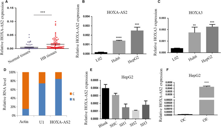
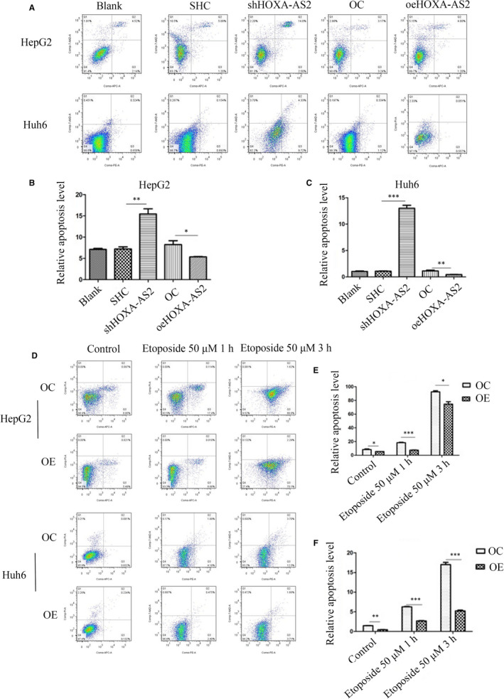
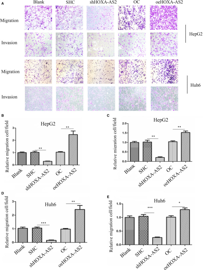
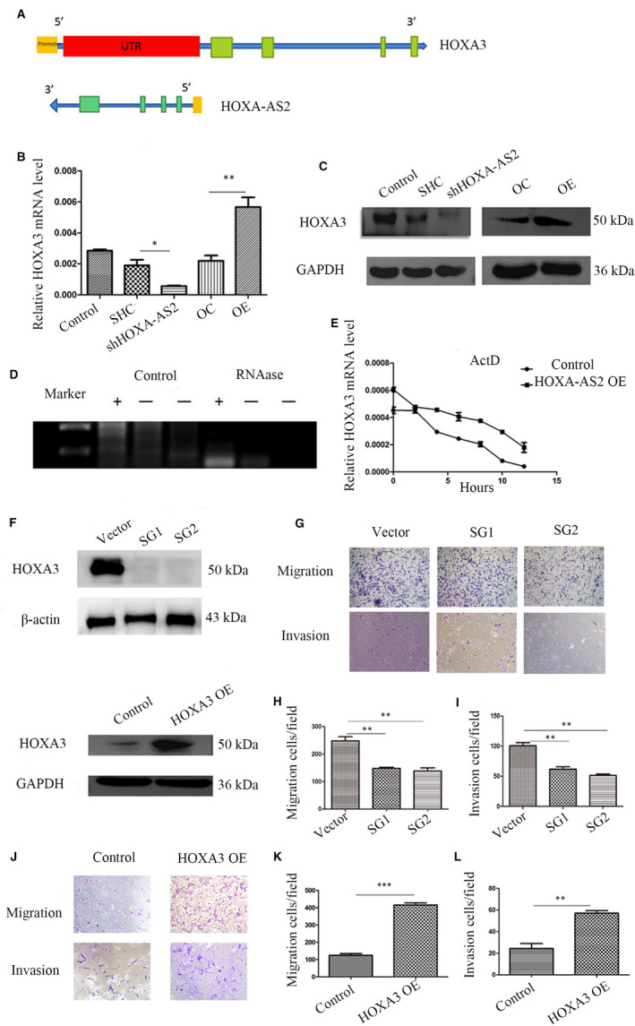
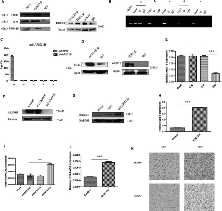
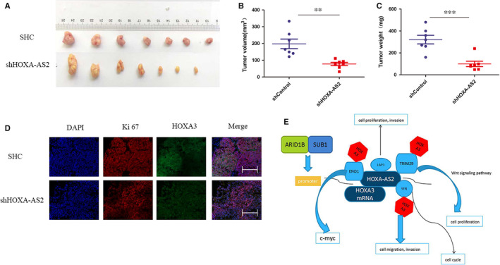
It has been becoming increasingly evident that long non-coding RNAs (lncRNAs) play important roles in various human cancers. However, the biological processes and clinical significance of most lncRNAs in hepatoblastoma (HB) remain unclear. In our previous study, genome-wide analysis with a lncRNA microarray found that lncRNA HOXA-AS2 was up-regulated in HB. Stable transfected cell lines with HOXA-AS2 knockdown or overexpression were constructed in HepG2 and Huh6 cells, respectively. Our data revealed knockdown of HOXA-AS2 increased cell apoptosis and inhibited cell proliferation, migration and invasion in HB. Up-regulation of HOXA-AS2 promoted HB malignant biological behaviours. Mechanistic investigations indicated that HOXA-AS2 was modulated by chromatin remodelling factor ARID1B and transcription co-activator SUB1, thereby protecting HOXA3 from degradation. Therefore, HOXA-AS2 positively regulates HOXA3, which might partly demonstrate the involvement of HOXA3 in HOXA-AS2-mediated HB carcinogenesis. In conclusion, HOXA-AS2 is significantly overexpressed in HB and the ARID1B/HOXA-AS2/HOXA3 axis plays a critical role in HB tumorigenesis and development. These results might provide a potential new target for HB diagnosis and therapy.

Keywords: chromatin remodelling factor; hepatoblastoma; lncRNA; tumorigenesis.


Conflict of interest statement

The authors declare that they have no conflict of interest.

Consent for publication: All the authors give their consent for publication.

Figures

FIGURE 1
FIGURE 1
LncRNA expression and localization. A, HOXA‐AS2 expression in the children HB and normal tissues was measured by RT‐PCR and relative to that of GAPDH; B, C, Expression of HOXA‐AS2 and HOXA3 in HepG2 cell, Huh6 cells and normal liver cells L02; D, Nuclear to cytoplasmic ratio of HOXA‐AS2 variants in HepG2 cells, analysed by RT‐qPCR. U1 was analysed as a nuclear control, and actin was analysed as a cytoplasmic control, respectively; E, F, the expression of HOXA‐AS2 in HepG2 transfected with shRNA or overexpression of HOXA‐AS2 or negative control which were evaluated by RT‐PCR. Blank represents no treatment, SHC represents empty vectors for shRNA, while SH1, SH2 and SH3 represent different targets for knockdown of HOXA‐AS2, OC represents empty vector for overexpression, and OE represents overexpression of HOXA‐AS2. Data are presented as the average and SD of three independent experiments (**P < .01,***P < .001 and ****<.0001)
FIGURE 2
FIGURE 2
HOXA‐AS2 was an apoptosis suppressor. A, B, C Determination of the apoptosis rate of HepG2 and Huh6 cells in each group by flow cytometry; D, E, F, Flow cytometry were used to detect HepG2 and Huh6 cell apoptosis with 50 μm etoposide for 1 h or 3 h. Blank represents no treatment, SHC represents empty vector for shRNA, shRNA represents knockdown of HOXA‐AS2, OC represents empty vector for overexpression, and OE represents overexpression of HOXA‐AS2. Data shown represent the average and SD of three independent experiments. *P < .05; **P < .01; ***P < .001
FIGURE 3
FIGURE 3
HOXA‐AS2 affected HB cell proliferation. A, B, Cell‐Light EdU DNA assay was used to reflect cell proliferation, the scale bars were 200 μm, and EdU positive represents cells that were proliferating; C, D, determination of HOXA‐AS2 silence or overexpression on the proliferation activity of HepG2 cells by CCK8 assay; E, F, determination of HOXA‐AS2 silence or overexpression on the proliferation activity of Huh6 cells by CCK8 assay; G, H, I, the colony formation of HepG2 and Huh6 cells determined by colony formation assay cell. Data shown represent the average and SD of three independent experiments. *P < .05; **P < .01; ***P < .001
FIGURE 4
FIGURE 4
Silencing of HOXA‐AS2 expression inhibited HB cells migration and invasion. A, Transwell assays demonstrating the effect of HOXA‐AS2 down‐regulation or up‐regulation on migration and invasion in HepG2 and Huh6 cells, the microscope magnification is 100×; B, C, statistics on the relative number of migration and invasion cells in HepG2; D, E, statistics on the relative number of migration and invasion cells in Huh6. Data shown represent the average and SD of three independent experiments. *P < .05; **P < .01; ***P < .001
FIGURE 5
FIGURE 5
HOXA‐AS2 regulated HOXA3 expression. A, Pattern diagram showing HOXA‐AS2 and HOXA3 mRNA binding; B, qRT‐PCR for detecting HOXA3 level with HOXA‐AS2 knockdown or overexpression; C, Western blot for detecting HOXA3 level with down‐regulation or up‐regulation of HOXA‐AS2; D, ribonuclease protection assay, ‘+’ represents the region of HOXA‐AS2 binding with HOXA3 mRNA, ‘−’ represents no binding region, control group represents without RNAase, RNAase group represents adding RNAase; E, actinomycin D (1 μm) was added in HOXA‐AS2 overexpression and control HepG2 cell at 0, 2, 4, 6, 8, 10 and 12 h, respectively, in mRNA stability test; F, Western blot for detecting HOXA3 knockdown or overexpression efficiency by CRISPR/CAS9; G,H,I Transwell assays demonstrating the effect of HOXA3 knockout on migration and invasion in HepG2 cells, the microscope magnification is 100×; J, K, L, migration and invasion capacity in HepG2 cells with HOXA3 overexpression, the microscope magnification is 100×. Data shown represent the average and SD of three independent experiments. *P < .05; **P < .01; ***P < .001
FIGURE 6
FIGURE 6
ARID1B and SUB1 co‐regulated HOXA‐AS2 expression. A, CO‐IP for HOXA3 and ENO1, SFN, TRIM29; B, C, CHIP‐PCR/QPCR for ARID1B and HOXA‐AS2 promoter(upstream of the transcription start site 2000 bp), a 1‐400 bp, b 400‐800 bp, c 800‐1200 bp, d 1200‐1600 bp, e 1600‐2000 bp, D, CO‐IP for ARID1B and SUB1, E, F, RT‐PCR and Western blot for detecting efficiency by down‐regulation of ARID1B; G, Western blot for HOXA3 with down‐regulation of ARID1B; H, J, RT‐PCR showing SUB1 overexpression efficiency and accompanying expression of HOXA‐AS2; I, RT‐PCR for HOXA‐AS2 with down‐regulation of ARID1B; K, immunohistochemistry for ARID1B and HOXA3 in HB tissues and adjacent normal liver tissues, the scale bars were 200 μm. Data shown represent the average and SD of three independent experiments. *P < .05; **P < .01; ***P < .001
FIGURE 7
FIGURE 7
HOXA‐AS2 regulated HB cell proliferation in vivo. A, The down‐regulation of HOXA‐AS2 inhibited the tumorigenesis ability of HB cells in mice, and nude mice were randomly divided into down‐regulation of HOXA‐AS2 group and control group (n = 7); B, the tumour volume of the nude mice in HOXA‐AS2 down‐regulation group and control group; C, the tumour weight of the nude mice in HOXA‐AS2 down‐regulation group and control group; D, immunofluorescence for Ki67 and HOXA3, and the scale bars were 200 μm; E, molecular mechanism diagram we envision. **P < .01; ***P < .001

Similar articles

Cited by

References

    1. Naoe A, Tsuchiya T, Kondo Y, et al. Arctigenin induces apoptosis in human hepatoblastoma cells. Pediatr Surg Int. 2019;35:723‐728. - PubMed
    1. Chowdappa R, Sagar RC, Ramesh S, Appaji L, Maneya P, Mukundapai M. Central hepatectomy (mesohepatectomy) by double liver hanging maneuver (DLHM) in a child with hepatoblastoma. Indian J Surg Oncol. 2019;10:46‐49. - PMC - PubMed
    1. Ofoegbu BN, Abdel Salam SEE, Diehl WG, Ghosn L. Congenital hepatoblastoma in a growing health economy. BMJ Case Rep. 2019;12:e223344. - PMC - PubMed
    1. Vishnoi JR, Sasidhar A, Misra S, et al. Hepatoblastoma in a young adult: a rare case report and review of the literature. J Gastrointest Cancer. 2019;51(1):319‐324. - PubMed
    1. Arcay A, Kintrup GT, Gelen MT, Arslan G, Karayalcin B. Hepatoblastoma mimicking hemangioma in labeled red blood cell scintigraphy. Clin Nucl Med. 2019;44:229‐231. - PubMed

Publication types

MeSH terms