Review
. 2021 Jun 17:72:17-46.
doi: 10.1146/annurev-arplant-081720-015238. Epub 2021 Mar 8.

Biological Phase Separation and Biomolecular Condensates in Plants

Affiliations
Review

Biological Phase Separation and Biomolecular Condensates in Plants

Ryan J Emenecker et al. Annu Rev Plant Biol. .

Abstract

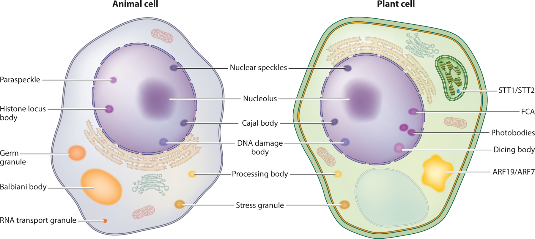
A surge in research focused on understanding the physical principles governing the formation, properties, and function of membraneless compartments has occurred over the past decade. Compartments such as the nucleolus, stress granules, and nuclear speckles have been designated as biomolecular condensates to describe their shared property of spatially concentrating biomolecules. Although this research has historically been carried out in animal and fungal systems, recent work has begun to explore whether these same principles are relevant in plants. Effectively understanding and studying biomolecular condensates require interdisciplinary expertise that spans cell biology, biochemistry, and condensed matter physics and biophysics. As such, some involved concepts may be unfamiliar to any given individual. This review focuses on introducing concepts essential to the study of biomolecular condensates and phase separation for biologists seeking to carry out research in this area and further examines aspects of biomolecular condensates that are relevant to plant systems.

Keywords: biomolecular condensates; membraneless organelles; protein phase separation.


Figures

Figure 1
Figure 1
Plant and animal cells contain many biomolecular condensates. A simplified animal cell (left) and plant cell (right) with various biomolecular condensates are depicted. Notably, some condensates appear to be conserved between plants and animals (terms between the two cells), whereas others appear to be specific to either animals (terms to the left of the animal cell) or plants (terms to the right of the plant cell). Abbreviations: ARF, AUXIN RESPONSE FACTOR; FCA, FLOWERING CONTROL LOCUS A.
Figure 2
Figure 2
Liquid-liquid phase separation (LLPS) of a simplified in vitro pseudo-two-component system. (a) LLPS is dependent on protein concentration, c (x axis), and other factors such as salt concentration; pH; or, as depicted here, temperature (y axis). At a fixed temperature (denoted by the horizontal line in the graph), as protein concentration increases from a relatively low concentration (c1) to a higher concentration (any concentration greater than c2 in this example), the protein will go from being homogeneously distributed throughout the system (the white one-phase regime) to existing in a two-phase regime (blue). In the two-phase regime, a protein-poor dilute phase and a protein-rich dense phase coexist with one another (❶ to ❷). In this example, c2 corresponds to the saturation concentration at the specified temperature associated with the horizontal line. In the two-phase regime, as overall protein concentration increases past c2, additional protein will be recruited into the dense phase of the system, resulting in an increase in the volume of the dense phase and a corresponding decrease in the volume of the dilute phase (❷ to ❸), Importantly, the protein concentration in the dense phase (c3) and dilute phase (c2) remains constant. As the total protein concentration increases further and while still in the two-phase regime, it will become more energetically favorable for the system to undergo an inversion transition, whereby dilute droplets exist in a majority dense phase (❸ to ❹). Finally, as protein concentration increases further, the system will exit the two-phase regime, exceeding the dense phase concentration (c3) such that the system will once again be homogeneously distributed in a one-phase regime. Once reentrance into the one-phase regime has occurred, the total concentration can continue to increase and exceed the concentration observed inside the dense-phase droplet (➎). (b) Phase diagrams built by changing different parameters can result in tie-lines having different slopes. Tie-lines are generated by connecting the solution conditions of the dense phase to the dilute phase. (i) For a tie-line generated in a system in which salt concentration is altered across the phase diagram, the concentration of salt in the dense phase may be different from that in the dilute phase, resulting in a tie-line that is not horizontal. (ii) In contrast, when temperature is altered, the temperature within the dilute and dense phases is expected to be equal, allowing for generation of a horizontal tie-line.
Figure 3
Figure 3
Multivalency can come in many different forms. Individual protein molecules can encode multivalency through various ways. (a) Linear multivalent molecules contain regions that facilitate inter- and intramolecular interactions (depicted as rectangular binding domains) connected by linkers that do not participate in multivalent interactions (linker regions). (b) Folded proteins can encode multivalency through many distinct interaction patches that facilitate multivalent interactions. (c) Intrinsically disordered proteins (or intrinsically disordered regions within a protein) can contain regions that facilitate multivalent interactions (stickers) separated by regions that do not participate in multivalent interactions (spacers). (d) Multiple individual proteins that are not multivalent on their own can come together through a central oligomerization domain, resulting in formation of a branched multivalent protein complex that can encode multivalency. (e) Oligomerization domains can contribute to phase separation both by contributing to multivalency and by bringing proteins within proximity of each other, increasing the likelihood of additional multivalent interactions such that oligomerization is a prerequisite for phase separation.
Figure 4
Figure 4
Examples of phase separation in plant biology. Plants use phase separation in the regulation of numerous fundamental plant growth and developmental processes. Nuclei are in purple; proteins of interest in blue. Nuclei appear blue when the protein of interest is diffusely in the nucleus. (a) FLOWERING CONTROL LOCUS A (FCA) forms subnuclear condensates and has been associated with regulation of flowering time in Arabidopsis (35). FLX-LIKE 2 (FLL2) (AT1G67170) regulates FCA condensate formation, and a recent study identified an FLL2 mutant that resulted in reduced FCA condensate formation and that had a delayed flowering phenotype, implicating FCA condensate formation in the regulation of flowering time (35). (b) NONEXPRESSOR OF PATHOGENESIS-RELATED GENES 1 (NPR1) is involved in regulation of effector-triggered immunity and forms oligomers in the cytoplasm through intramolecular disulfide bonds when there is no oxidative stress (125). However, upon pathogen attack, intracellular redox changes result in an oligomer-to-monomer transition for NPR1, resulting in dispersion of cytoplasmic oligomers (125). (c) STT1 (At5G40160) and STT2 (At5G66055) mediate translocation of proteins in the chloroplast twin-arginine translocation (cpTat) pathway from the stroma to the thylakoid membranes via a phase separation–dependent mechanism (91). STT1 and STT2 form condensates within the chloroplast, and disruption of STT1 and STT2 phase separation results in disruption of cpTat substrate translocation across the stroma (91). (d) AUXIN RESPONSE FACTOR (ARF)7 and ARF19 are localized to the nucleus near the root tip but are localized to cytoplasmic condensates in the upper root, where cell growth has ceased (102). The localization of ARF7/ARF19 to cytoplasmic condensates attenuates auxin response in the upper root and is important for regulation of auxin responsiveness in the root (102). (e) PhyB photobodies are subnuclear condensates that form in response to red light (52). PhyB photobodies have been implicated in the regulation of the circadian clock, photomorphogenesis, and thermomorphogenesis (52). Abbreviations: C, chloroplast; N, nucleus.

References

    1. Alberti S, Dormann D. 2019. Liquid-liquid phase separation in disease. Annu. Rev. Genet 53:171–94 - PubMed
    1. Alberti S, Gladfelter A, Mittag T. 2019. Considerations and challenges in studying liquid-liquid phase separation and biomolecular condensates. Cell 176:419–34 - PMC - PubMed
    1. Alberti S, Saha S, Woodruff JB, Franzmann TM, Wang J, Hyman AA. 2018. A user’s guide for phase separation assays with purified proteins. J. Mol. Biol 430:4806–20 - PMC - PubMed
    1. Banani SF, Lee HO, Hyman AA, Rosen MK. 2017. Biomolecular condensates: organizers of cellular biochemistry. Nat. Rev. Mol. Cell Biol 18:285–98 - PMC - PubMed
    2. Comprehensively reviews biomolecular condensates.

    1. Banani SF, Rice AM, Peeples WB, Lin Y, Jain S, et al. 2016. Compositional control of phase-separated cellular bodies. Cell 166:651–63 - PMC - PubMed

Publication types

LinkOut - more resources