Review
. 2021 Sep 1;94(1125):20201332.
doi: 10.1259/bjr.20201332. Epub 2021 May 5.

Differentiating uterine sarcoma from leiomyoma: BET1T2ER Check!

Affiliations
Review

Differentiating uterine sarcoma from leiomyoma: BET1T2ER Check!

Janette Smith et al. Br J Radiol. .

Abstract

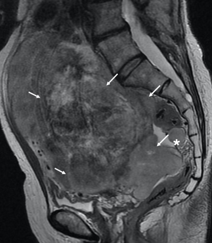
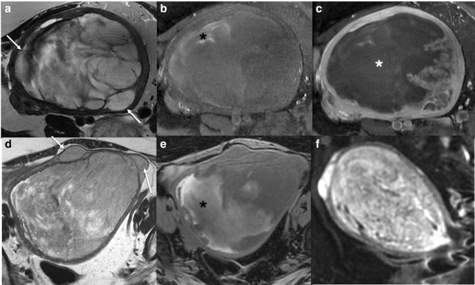
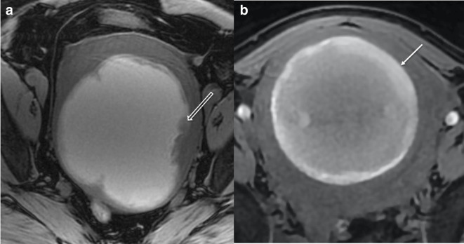
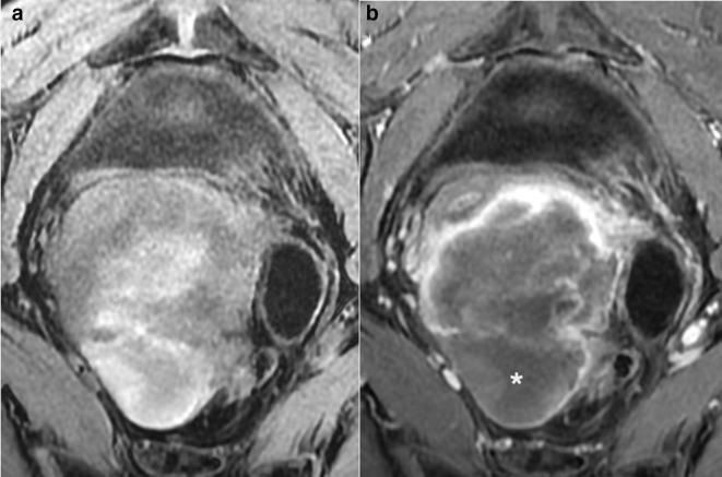
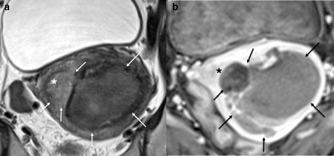
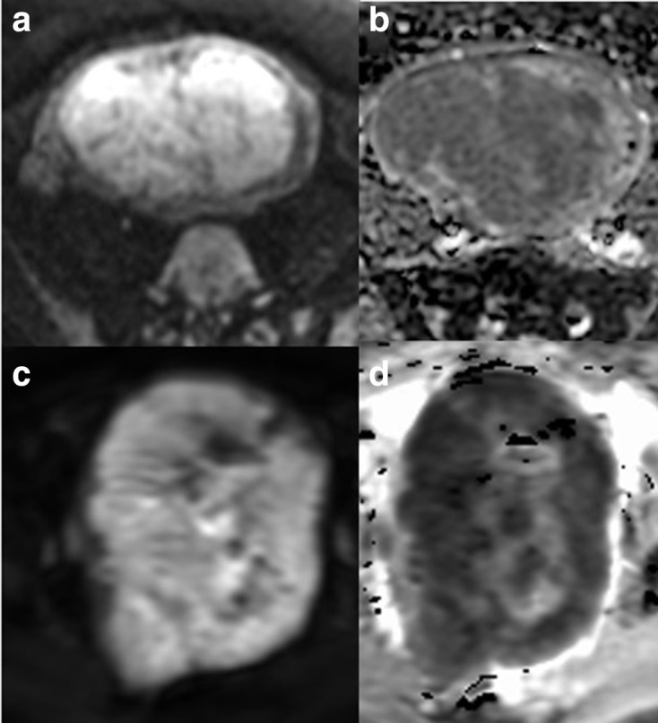
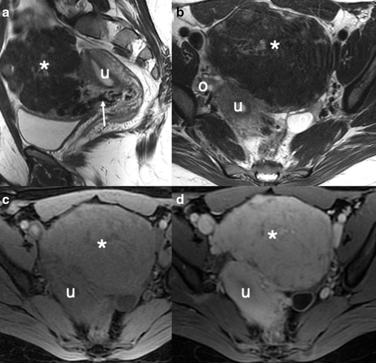
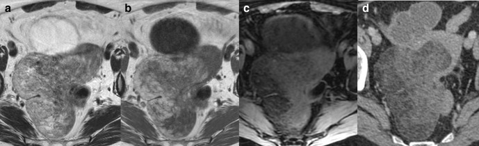
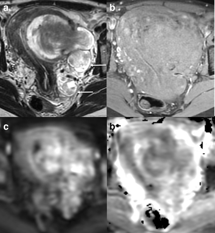
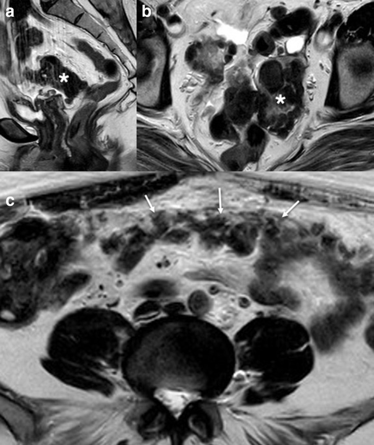
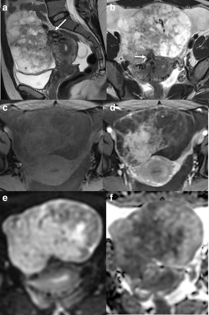
Although rare, uterine sarcoma is a diagnosis that no one wants to miss. Often benign leiomyomas (fibroids) and uterine sarcomas can be differentiated due to the typical low T2 signal intensity contents and well-defined appearances of benign leiomyomas compared to the suspicious appearances of sarcomas presenting as large uterine masses with irregular outlines and intermediate T2 signal intensity together with possible features of secondary spread. The problem is when these benign lesions are atypical causing suspicious imaging features. This article provides a review of the current literature on imaging features of atypical fibroids and uterine sarcomas with an aide-memoire BET1T2ER Check! to help identify key features more suggestive of a uterine sarcoma.


Figures

Figure 1.
Figure 1.
Sagittal T2W MRI of a spindle cell leiomyosarcoma demonstrating aggressive features such as a heterogenous T2W intrauterine lesion, irregular border extending through the uterine serosa (white arrows) and additional separate extra uterine peritoneal deposit (*).
Figure 2.
Figure 2.
Axial T2 (a) and T1 fat saturated image without (b) and with gadolinium contrast medium (c) MRI of a low grade ESS. The ESS demonstrates myometrial invasion with irregular nodular borders (a, white arrows), heterogenous T2 signal, intralesional haemorrhage (b, black *), and central poor enhancement (c, white *). Axial T2W (d), axial T1 fat saturated image (e) and DWI (f) MRI of a uterine adenosarcoma. The adenosarcoma demonstrates myometrial invasion with irregular borders (d, white arrows), heterogenous T2 signal, intralesional haemorrhage (e, black *), and restricted diffusion. (f). ADC maps not shown. ADC, apparent diffusioncoefficient; DWI, diffusion-weighted imaging; ESS, endometrial stromal sarcoma.
Figure 3.
Figure 3.
Comparison of imaging in LMS (a) and red cell degeneration of benign fibroid (b): Axial T1W fat saturated MRI of two intrauterine lesions. Lesion (a) has diffuse hyperintense T1W fat saturated signal with and irregular intermediate T1 signal border and soft tissue projecting into the lesion (open white arrow). Lesion (b) has an intermediate to high T1W fat saturated signal intensity with a hyperintense smooth rim and clear distinction of the margin with no internal projections (closed white arrow) in a 40-year-old lady. Surgical histology of lesion (a) confirmed LMS. Lesion (b) had been imaged with MRI 12 months previously, when it had low T2W signal. Between the two MRIs, the female had become pregnant and given birth and the follow-up MRI was for further management of her benign leiomyoma, which had undergone red degeneration during pregnancy. LMS, leiomyosarcoma.
Figure 4.
Figure 4.
Axial T1W fat saturated image without (a) and with (b) gadolinium contrast medium. (a) demonstrates high T1 fat sat signal intensity in keeping with intralesional haemorrhage. Central unenhanced areas are seen in (b) in keeping with a high grade sarcoma (confirmed on histology).
Figure 5.
Figure 5.
Sagittal (a) and axial (b) T2W MRI of a high grade leiomyosarcoma demonstrating heterogenous T2 signal intensity with focal hypointense T2 areas (*) in keeping with haemosiderin within the intrauterine lesion.
Figure 6.
Figure 6.
Axial T2W (a) and axial T1 fat saturated gadolinium contrast medium enhanced (b) MRI of a high grade leiomyosarcoma demonstrating endometrial invasion (*) by a leiomyosarcoma (arrows).
Figure 7.
Figure 7.
Comparison of DWI imaging in benign (a, b) and malignant (c, d) lesions: axial DWI (a) and ADC (b) of a highly cellular leiomyoma and axial DWI (c) and ADC (d) of a leiomyosarcoma. The DWI outline is helpful in demonstrating the irregular outline in the malignant lesion (c, d). ADC, apparent diffusioncoefficient; DWI, diffusion-weighted imaging.
Figure 8.
Figure 8.
Typical benign fibroid: sagittal (a) and axial (b) T2W images demonstrating a well-defined low SI mass (*) extending anteriorly from the uterus (u) and separate to the ovaries (o) with bridging vessels (white arrow) in keeping with a subserosal fibroid. The ‘whorled’ and low T2WI SI is typical for benign fibroid. Axial T1W images before (c) and following (d) the administration of intravenous contrast medium demonstrates intense enhancement of the fibroid with no necrosis.
Figure 9.
Figure 9.
Axial T2W (a), T1W (b) and T1W fat saturated (c) images of a uterine lipoleiomyoma. Axial portal venous contrast-enhanced CT of the pelvis (d) confirms fat attenuation within the uterine lesion.
Figure 10.
Figure 10.
Axial T2W (a), T1W fat saturated image (b), DWI (c) and ADC (d) of a uterine lesion in a 45-year-old females. Heterogenous T2W lesion demonstrates extension though the myometrium with high T2 signal ‘deposits’ within the left of the pelvis (white arrows). No lymph node enlargement or metastasis was seen on staging CT. Pre-operative diagnosis of a uterine sarcoma was made and a total hysterectomy and bilateral salpingo-oophrectomy performed. Leiomyomatosis with intravascular invasion was confirmed on histology. ADC, apparent diffusioncoefficient; DWI, diffusion-weighted imaging.
Figure 11.
Figure 11.
Sagittal (a) and axial (b) T2W images demonstrating previous hysterectomy (for fibroids) with a well-defined low signal intensity mass (*). Axial T2W (c) image of the abdomen demonstrates low signal intensity mass outlining the peritoneum (white arrows). This had been mistaken for an ovarian Stage 3C malignancy on CT imaging but the CA-125 level was normal. Biopsy of the peritoneal area confirmed peritoneal leiomyomatosis.
Figure 12.
Figure 12.
Sagittal (a) and axial (b) T2W images of an exophytic uterine lesion with a narrow base. The lesion demonstrates bridging vessels (White arrow) and cystic peripheral changes. The central area of enhancement and peripheral poor enhancement is seen on the T1 fat saturated imaging without (c) and with gadolinium contrast medium (d). DWI (e) and ADC (f) demonstrate central restricted diffusion within the lesion. Pre-operatively imaging suggested a torted large subserosal leiomyoma. However, on histology, a smooth muscle tumour of uncertain malignant potential was diagnosed. ADC, apparent diffusioncoefficient; DWI, diffusion-weighted imaging

References

    1. Wu T-I, Yen T-C, Lai C-H. Clinical presentation and diagnosis of uterine sarcoma, including imaging. Best Pract Res Clin Obstet Gynaecol 2011; 25: 681–9. doi: 10.1016/j.bpobgyn.2011.07.002 - DOI - PubMed
    1. Thomassin-Naggara I, Dechoux S, Bonneau C, Morel A, Rouzier R, Carette M-F, et al. . How to differentiate benign from malignant myometrial tumours using MR imaging. Eur Radiol 2013; 23: 2306–14. doi: 10.1007/s00330-013-2819-9 - DOI - PubMed
    1. Kubik-Huch RA, Weston M, Nougaret S, Leonhardt H, Thomassin-Naggara I, Horta M, et al. . European Society of urogenital radiology (ESUR) guidelines: MR imaging of leiomyomas. Eur Radiol 2018; 28: 3125–37. doi: 10.1007/s00330-017-5157-5 - DOI - PMC - PubMed
    1. Bi Q, Xiao Z, Lv F, Liu Y, Zou C, Shen Y. Utility of clinical parameters and multiparametric MRI as predictive factors for differentiating uterine sarcoma from atypical leiomyoma. Acad Radiol 2018; 25: 993–1002. doi: 10.1016/j.acra.2018.01.002 - DOI - PubMed
    1. Lakhman Y, Veeraraghavan H, Chaim J, Feier D, Goldman DA, Moskowitz CS, et al. . Differentiation of uterine leiomyosarcoma from atypical leiomyoma: diagnostic accuracy of qualitative MR imaging features and feasibility of texture analysis. Eur Radiol 2017; 27: 2903–15. doi: 10.1007/s00330-016-4623-9 - DOI - PMC - PubMed