Review
. 2021 May 1;94(1121):20201457.
doi: 10.1259/bjr.20201457. Epub 2021 Mar 18.

Atraumatic fractures of the femur

Affiliations
Review

Atraumatic fractures of the femur

Ganesh Hedge et al. Br J Radiol. .

Abstract

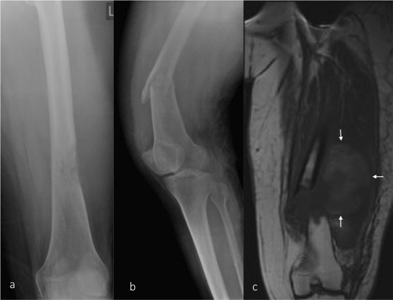
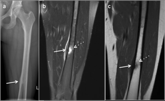
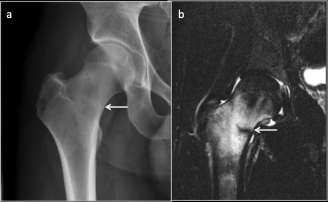
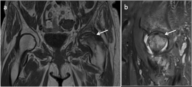
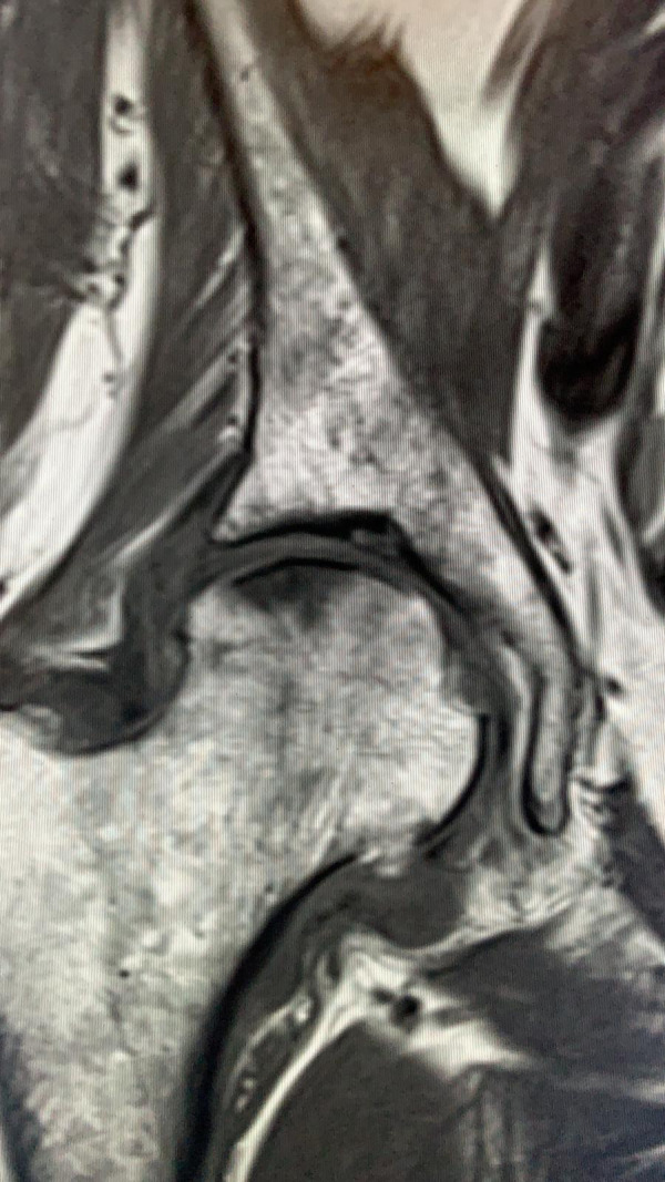
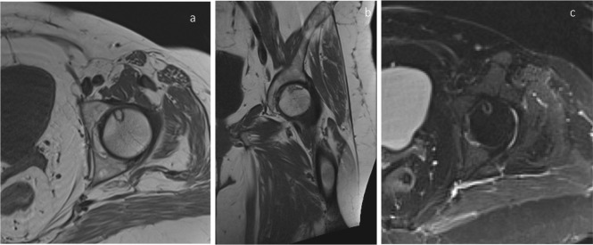
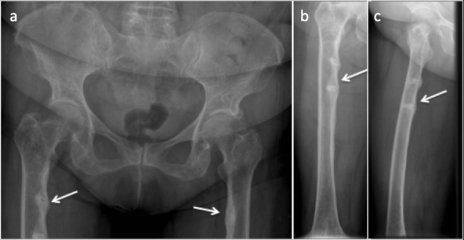
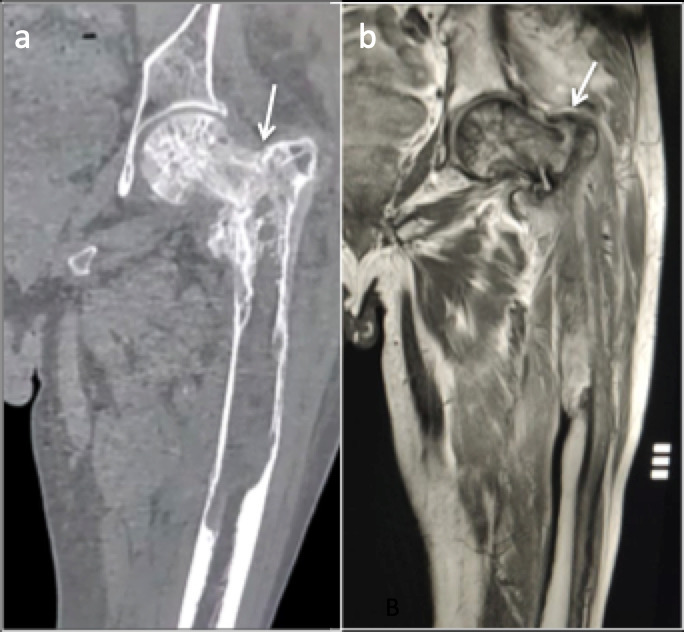
Atraumatic fractures of femur, although not as common as traumatic fractures, are frequently encountered in the clinical practice. They present with non-specific symptoms and can be occult on initial imaging making their diagnosis difficult, sometimes resulting in complications. Overlapping terminologies used to describe these fractures may hamper effective communication between the radiologist and the clinician. In this article, we review various atraumatic fractures of femur, terminologies used to describe them, their imaging findings and differential diagnosis. The article also describes the aetiology, pathophysiology and relevant biomechanics behind these fractures. An approach to atraumatic femoral fractures has been outlined.


Figures

Figure 1.
Figure 1.
Pathological fracture/ (a) AP radiograph of the femur demonstrating a lucent lesion in the distal femur with permeative pattern of bone destruction suggesting an aggressive lesion. (b) Lateral view of the knee joint including the distal femur demonstrating a pathological fracture through the lesion. (c) T1 coronal MRI image showing aggressive lesion replacing the normal marrow fat at the fracture site with a large adjacent soft tissue component. AP, anteroposterior.
Figure 2.
Figure 2.
35-year-old female amateur marathon runner with Fatigue fracture in the femoral diaphysis where cortical bone predominates. (a) Radiograph demonstrating subtle central lucency with cortical thickening and periosteal reaction (arrow). (b) Coronal PDFS image (c) Coronal T1 image demonstrating marrow edema (dotted arrow), cortical thickening and periosteal reaction (arrow). PDFS, proton density fat saturated.
Figure 3.
Figure 3.
Fatigue fracture in the femoral neck where cancellous bone predominates. (a) Radiograph demonstrating subtle intramedullary sclerosis (arrow) along the fracture line with blurred trabeculae. (b) Coronal STIR image demonstrating marrow oedema and the linear hypointense fracture line(arrow). STIR, short tau inversion recovery.
Figure 4.
Figure 4.
Subchondral insufficiency fracture of the left femoral head. Coronal T1 (a) and Sagittal PDFS (b) images demonstrating marrow oedema in the femoral head and neck with Hypo intense irregular fracture line (arrow). PDFS, proton density fat saturated.
Figure 5.
Figure 5.
MRI Coronal T1 imaging of right hip demonstrating a linear low signal subchondral line in the superior femoral head consistent with subchondral insufficiency fracture.
Figure 6.
Figure 6.
T1 axial (a) and Coronal (b) and T2 fat saturated axial (c) images demonstrating osteonecrosis (avascular necrosis) with a smooth well-circumscribed margin.
Figure 7.
Figure 7.
(a) Anteroposterior radiograph of the pelvis. (b) Anteroposterior and (c) Lateral radiograph of the right femur showing looser zones of osteomalacia. Multiple, bilateral lucency which involve only a part of femoral shaft along the medial aspect, oriented perpendicular to the cortex with associated surrounding sclerosis.
Figure 8.
Figure 8.
Paget’s disease of left proximal femur with slight bowing change and intertrochanteric fracture (arrows). (a) Coronal CT reformat and (b) Coronal MRI T1 imaging.
Figure 9.
Figure 9.
Anteroposterior radiograph of the proximal femur with atypical femoral fracture in the left proximal femur. Note the lucent transverse fracture line with endosteal and periosteal beaking (arrow).
Figure 10.
Figure 10.
Bilateral involvement of atypical femoral fracture. 73-year-old female on bisphosphonates (a) Complete displaced fracture of the right femoral shaft. (b) Cortical and periosteal thickening and beaking in the outer cortex of the left femoral shaft indicating early changes of incomplete atypical femoral fracture (arrow).
Figure 11.
Figure 11.
56 year-old-female with hypophosphatasia with atypical femoral fracture. Anteroposterior radiograph of the femur (a) demonstrates a lateral diaphysis lucency and MRI PDFS sequences (b, c) transverse cortical lucency (arrows) along the lateral femoral cortex. PDFS, proton density fat saturated.
Figure 12.
Figure 12.
Patient presenting with hip pain. (a) Radiograph demonstrating central lucency (arrow) with surrounding sclerosis and cortical thickening along the medial cortex of proximal femur resembling a fatigue fracture. (b) Coronal and (c) Axial CT images demonstrating central nidus and surrounding sclerosis and cortical thickening indicating that the lesion is osteoid osteoma.
Figure 13.
Figure 13.
Osteomyelitis with cortical abscess femur. Anteroposterior (a) radiograph of the proximal femur showing lucent lesion in the medial cortex (arrow) with adjacent periosteal reaction. Coronal T1(b), coronal and axial STIR (c, d) MRI images demonstrating intracortical abscess (arrow) with extensive marrow and adjacent soft tissue oedema. STIR, short tau inversion recovery.

Similar articles

Cited by

References

    1. Marshall RA, Mandell JC, Weaver MJ, Ferrone M, Sodickson A, Khurana B. Imaging features and management of stress, atypical, and pathologic fractures. Radiographics 2018; 38: 2173–92. doi: 10.1148/rg.2018180073 - DOI - PubMed
    1. Pentecost RL, Murray RA, Brindley HH. Fatigue, insufficiency, and pathologic fractures. JAMA 1964; 187: 1001-4. doi: 10.1001/jama.1964.03060260029006 - DOI - PubMed
    1. Anderson MW, Greenspan A. Stress fractures. Radiology 1996; 199: 1–12. doi: 10.1148/radiology.199.1.8633129 - DOI - PubMed
    1. Wagner D, Ossendorf C, Gruszka D, Hofmann A, Rommens PM. Fragility fractures of the sacrum: how to identify and when to treat surgically? Eur J Trauma Emerg Surg 2015; 41: 349–62. doi: 10.1007/s00068-015-0530-z - DOI - PMC - PubMed
    1. Belthur MV, Birchansky SB, Verdugo AA, Mason EO, Hulten KG, Kaplan SL, et al. . Pathologic fractures in children with acute Staphylococcus aureus osteomyelitis. J Bone Joint Surg Am 2012; 94: 34–42. doi: 10.2106/JBJS.J.01915 - DOI - PubMed