

In vivo brain imaging of mitochondrial Ca2+ in neurodegenerative diseases with multiphoton microscopy
- PMID: 33684410
- PMCID: PMC8057769
- DOI: 10.1016/j.bbamcr.2021.118998
In vivo brain imaging of mitochondrial Ca2+ in neurodegenerative diseases with multiphoton microscopy
Abstract
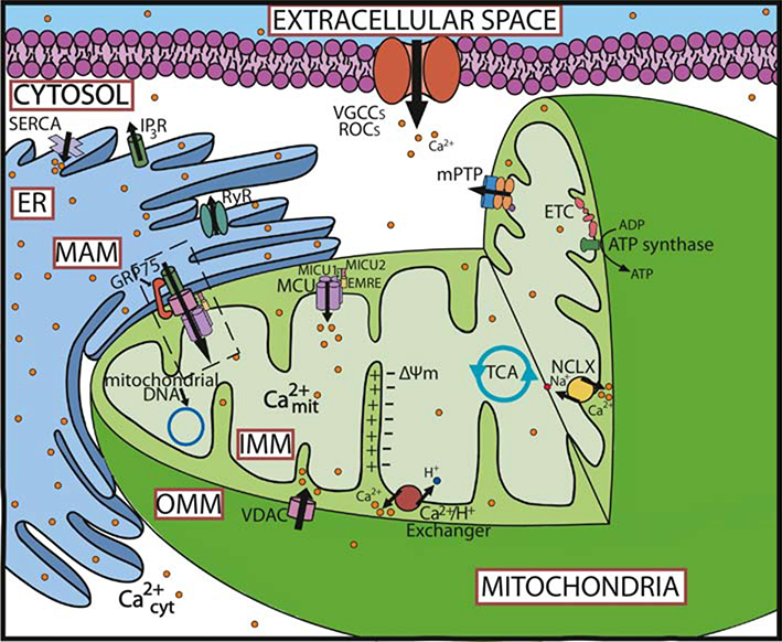
Mitochondria are involved in a large number of essential roles related to neuronal function. Ca2+ handling by mitochondria is critical for many of these functions, including energy production and cellular fate. Conversely, mitochondrial Ca2+ mishandling has been related to a variety of neurodegenerative diseases. Investigating mitochondrial Ca2+ dynamics is essential for advancing our understanding of the role of intracellular mitochondrial Ca2+ signals in physiology and pathology. Improved Ca2+ indicators, and the ability to target them to different cells and compartments, have emerged as useful tools for analysis of Ca2+ signals in living organisms. Combined with state-of-the-art techniques such as multiphoton microscopy, they allow for the study of mitochondrial Ca2+ dynamics in vivo in mouse models of the disease. Here, we provide an overview of the Ca2+ transporters/ion channels in mitochondrial membranes, and the involvement of mitochondrial Ca2+ in neurodegenerative diseases followed by a summary of the main tools available to evaluate mitochondrial Ca2+ dynamics in vivo using the aforementioned technique.
Keywords: Alzheimer's disease; Calcium; Fluorescent proteins; GECIs; Mitochondria; Multiphoton microscopy.
Copyright © 2021. Published by Elsevier B.V.
Conflict of interest statement
Declaration of interests
The authors declare that they have no known competing financial interests or personal relationships that could have appeared to influence the work reported in this paper.
Conflict of interest
The authors declare no competing interests.
Figures
Similar articles
-
The mitochondrial calcium signaling, regulation, and cellular functions: A novel target for therapeutic medicine in neurological disorders.J Cell Biochem. 2023 May;124(5):635-655. doi: 10.1002/jcb.30414. Epub 2023 May 9. J Cell Biochem. 2023. PMID: 37158125 Review.
-
Calcium dysregulation and homeostasis of neural calcium in the molecular mechanisms of neurodegenerative diseases provide multiple targets for neuroprotection.Antioxid Redox Signal. 2011 Apr 1;14(7):1275-88. doi: 10.1089/ars.2010.3359. Epub 2010 Oct 6. Antioxid Redox Signal. 2011. PMID: 20615073 Free PMC article. Review.
-
Increased mitochondrial calcium levels associated with neuronal death in a mouse model of Alzheimer's disease.Nat Commun. 2020 May 1;11(1):2146. doi: 10.1038/s41467-020-16074-2. Nat Commun. 2020. PMID: 32358564 Free PMC article.
-
Overexpression of Mitochondrial Calcium Uniporter Causes Neuronal Death.Oxid Med Cell Longev. 2019 Oct 16;2019:1681254. doi: 10.1155/2019/1681254. eCollection 2019. Oxid Med Cell Longev. 2019. PMID: 31737163 Free PMC article.
-
Mitochondrial Ca(2+) and neurodegeneration.Cell Calcium. 2012 Jul;52(1):73-85. doi: 10.1016/j.ceca.2012.04.015. Epub 2012 May 18. Cell Calcium. 2012. PMID: 22608276 Free PMC article. Review.
Cited by
-
Calcium imaging: A versatile tool to examine Huntington's disease mechanisms and progression.Front Neurosci. 2022 Nov 3;16:1040113. doi: 10.3389/fnins.2022.1040113. eCollection 2022. Front Neurosci. 2022. PMID: 36408400 Free PMC article. Review.
-
Ultrafast optical imaging techniques for exploring rapid neuronal dynamics.Neurophotonics. 2025 Jan;12(Suppl 1):S14608. doi: 10.1117/1.NPh.12.S1.S14608. Epub 2025 Feb 27. Neurophotonics. 2025. PMID: 40017464 Free PMC article. Review.
-
Updated Toolbox for Assessing Neuronal Network Reconstruction after Cell Therapy.Bioengineering (Basel). 2024 May 14;11(5):487. doi: 10.3390/bioengineering11050487. Bioengineering (Basel). 2024. PMID: 38790353 Free PMC article. Review.
-
Genetically encoded biosensors of metabolic function for the study of neurodegeneration, a review and perspective.Neurophotonics. 2025 Jun;12(Suppl 2):S22805. doi: 10.1117/1.NPh.12.S2.S22805. Epub 2025 Sep 4. Neurophotonics. 2025. PMID: 40919264 Free PMC article. Review.
-
The Role of Oxygen Homeostasis and the HIF-1 Factor in the Development of Neurodegeneration.Int J Mol Sci. 2024 Apr 23;25(9):4581. doi: 10.3390/ijms25094581. Int J Mol Sci. 2024. PMID: 38731800 Free PMC article. Review.
References
-
- Attwell D, Laughlin SB, An energy budget for signaling in the grey matter of the brain, Journal of cerebral blood flow and metabolism : official journal of the International Society of Cerebral Blood Flow and Metabolism, 21 (2001) 1133–1145. - PubMed
Publication types
MeSH terms
Substances
Grants and funding
LinkOut - more resources
Full Text Sources
Other Literature Sources
Medical
Miscellaneous
