Review
. 2021 Jan-Jun:296:100521.
doi: 10.1016/j.jbc.2021.100521. Epub 2021 Mar 5.

RHO to the DOCK for GDP disembarking: Structural insights into the DOCK GTPase nucleotide exchange factors

Affiliations
Review

RHO to the DOCK for GDP disembarking: Structural insights into the DOCK GTPase nucleotide exchange factors

Andrew P Thompson et al. J Biol Chem. 2021 Jan-Jun.

Abstract

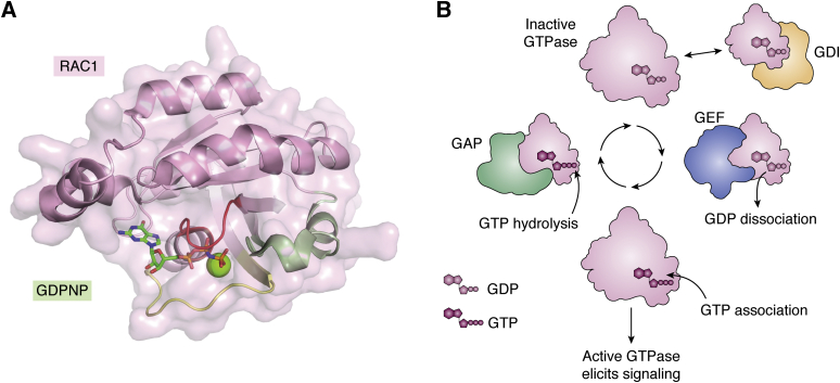
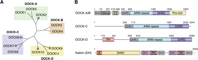
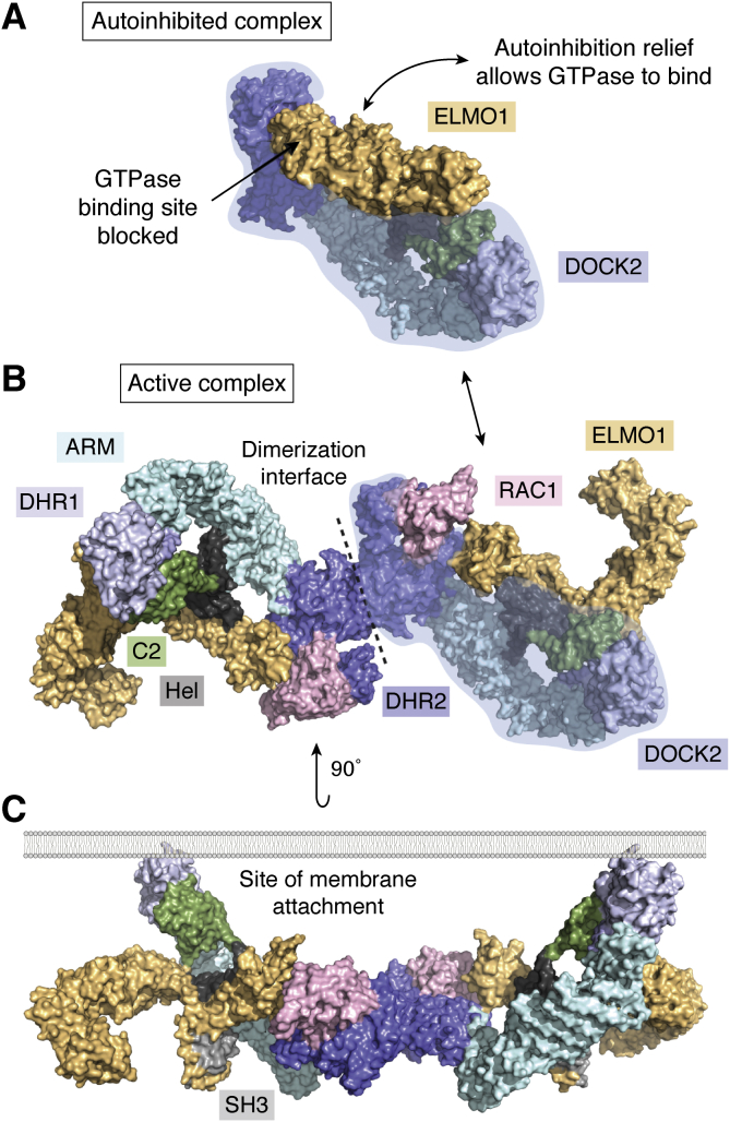
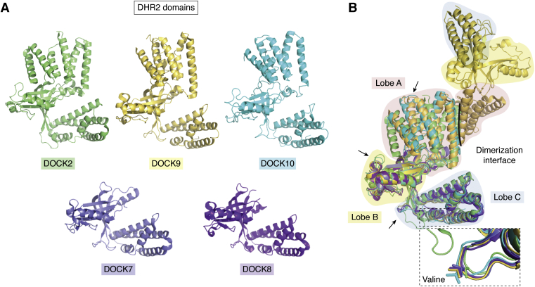
The human dedicator of cytokinesis (DOCK) family consists of 11 structurally conserved proteins that serve as atypical RHO guanine nucleotide exchange factors (RHO GEFs). These regulatory proteins act as mediators in numerous cellular cascades that promote cytoskeletal remodeling, playing roles in various crucial processes such as differentiation, migration, polarization, and axon growth in neurons. At the molecular level, DOCK DHR2 domains facilitate nucleotide dissociation from small GTPases, a process that is otherwise too slow for rapid spatiotemporal control of cellular signaling. Here, we provide an overview of the biological and structural characteristics for the various DOCK proteins and describe how they differ from other RHO GEFs and between DOCK subfamilies. The expression of the family varies depending on cell or tissue type, and they are consequently implicated in a broad range of disease phenotypes, particularly in the brain. A growing body of available structural information reveals the mechanism by which the catalytic DHR2 domain elicits nucleotide dissociation and also indicates strategies for the discovery and design of high-affinity small-molecule inhibitors. Such compounds could serve as chemical probes to interrogate the cellular function and provide starting points for drug discovery of this important class of enzymes.

Keywords: Ras homologous (RHO) small GTPases; cell signaling; dedicator of cytokinesis (DOCK); drug discovery; guanine nucleotide exchange factor; guanosine triphosphate (GTP); structural biology.


Conflict of interest statement

Conflict of interest The authors declare that they have no conflicts of interest with the contents of this article.

Figures

Figure 1
Figure 1
A, RHO family small GTPase RAC1 (pink surface, cartoon; PDB ID: 3TH5) bound to phosphoaminophosphonic acid–guanylate ester (GDPNP; green sticks) and magnesium (green sphere). The phosphate-binding loop (P-loop; red ribbon; residues 10–17) binds the phosphates of guanosine polyphosphates and magnesium. Switch loops 1 and 2 (yellow and green ribbons; residues 27–40, 57–74 respectively) change conformation depending on the presence of GDP or GTP to affect cellular signaling. B, GTPases (pink) cycle between an inactive, GDP-bound state to an active, GTP-bound state. The switch loop conformations in the active state allow GTPases to bind to and elicit cellular signaling processes. GDP dissociation is prevented by guanosine dissociation inhibitors (GDIs, orange) and accelerated by guanosine nucleotide exchange factors (GEFs, blue), while GTP hydrolysis is induced by GTPase-activating proteins (GAPs). Together these interactions tightly control the location and timing of GTPase activity. Figure created with BioRender.com.
Figure 2
Figure 2
A, DOCK proteins are largely classified into their subfamilies based on phylogeny, as well as sequence and substrate specificity. DOCK proteins with publicly available structural information are indicated by a circle. B, domain architecture of the DOCK proteins also follows their subfamilial categorization. All DOCK proteins contain a DHR1 as well as the catalytic DHR2 domain. Domains and numbering for the DOCK-A and B are based on the recently published Cryo-EM structure of the full-length DOCK2. The ARM repeat is putatively present in all DOCK proteins based on structural homology prediction. Domain numbering is represented by DOCK6 and 9 for DOCK-C and D subfamilies respectively. The domain architecture for Kalirin is provided as an example Dbl-Homology GEF. Kalirin is truncated for clarity of comparison (represented by dashed line). The canonical DH/PH architecture present twice in Kalirin confers its GEF activities.
Figure 3
Figure 3
A, cryo-EM structure of the autoinhibited DOCK2-ELMO1 complex (PDB ID: 6TGC). The DOCK2 (monomer depicted by transparent blue highlight) GTPase-binding site is occupied by ELMO1 (yellow). B, cryo-EM structure of the active DOCK2-ELMO-RAC1 complex (PDB ID: 6TGC). DOCK2 dimerizes through the DHR2 domain (blue), which also binds RAC1 (pink). ELMO1 binds to the SH3, Helical (Hel, black), C2 (green), and DHR2 domains, as well as the C-terminal pro-rich sequence (not depicted). ELMO1 undergoes conformational change upon relief of the autoinhibitory state depicted in panel A. C, the entire complex is localized to the membrane through the phospholipid-binding capabilities of the DOCK2 DHR1 and C2 domains. Complex is rotated 90 degrees out of the page with respect to B.
Figure 4
Figure 4
A, the individual DOCK DHR2 domains, represented as monomers.B, superposition of the DOCK DHR2 domains illustrates their similar overall fold that is segregated into three lobes A–C. Lobe A is made of 5 to 6 alpha helices depending on subfamily, and variations are observed in the interjoining loops between helices (examples highlighted by arrows). This lobe is the site of homodimerization (only DOCK9 dimer shown). Lobe A is not present in the structures of DOCK7 and 8 although predicted to be present based on sequence homology. Lobes B and C are responsible for GTPase substrate binding, discrimination, and catalytic activity. Variability is also observed in the interjoining loops between secondary structures (examples highlighted by arrows). Lobe C contains a universally conserved valine on the α10 insert (highlighted by dotted box), which is responsible for occluding the magnesium that is crucial for the GDP binding. DOCK2 PDB ID: 2YIN; DOCK7 PDB ID: 6AJ4; DOCK8 PDB ID: 3VHL; DOCK9 PDB ID: 2WM9; DOCK10 PDB ID: 6TM1.
Figure 5
Figure 5
A, GDP (green sticks) binds to CDC42 (cyan surface, sticks) through an extensive network of intermolecular interactions. Hydrogen bonding and salt bridges are indicated by dashed yellow lines. Most interactions are concentrated around the nucleoside-binding region (cyan sticks) and the phosphate-binding loop (P-loop; red sticks). Right, crucial CDC42 interactions in the GEF -induced GDP dissociation mechanism are highlighted: 1. Phe28 is located in the switch 1 (yellow cartoon, sticks); 2. Cys18 is proximal to the P-loop; 3. Mg2+ (small green sphere) is chelated by GDP phosphates, the P-loop, and several water molecules (small red spheres). (CDC42 PDB ID: 1A4R). B, Left, DOCK9 (Orange surface and cartoon) binds to GDP-bound CDC42 (blue surface) via lobes B and C. For orientation, this representation is the underside of the view presented in Figure 4. Right, close-up of the CDC42 GTP-binding site when bound to DOCK9. Only the critical features involved in the GDP dissociation mechanism are presented. Phe28 and Cys18 rotate away from the GDP molecule, while the DOCK9-Val1951 juts into the active site and occludes Mg2+ binding. The dotted sphere indicates where Mg2+ binding was before DOCK9 binding. The cumulative result of these conformational changes leads to significantly decreased affinity between CDC42 and GDP. (DOCK9:CDC42 PDB ID: 2WMN). Notably, the switch 2 region (green ribbon) of CDC42 is unchanged on DOCK binding.
Figure 6
Figure 6
Potential options for drugging the DOCK-GTPase complex.Left, overview of the DOCK2-RAC1 complex (electrostatic surface, pink ribbon respectively; PDB ID: 2YIN). A, an electrostatic representation of the DOCK2-RAC1 interface, overlaid with RAC1-GDPNP (PDB ID: 3TH5) to visualize the GTP (green sticks) binding site. The RAC1 surface is highlighted by the transparent white mask. The nucleotide sensing Val1539 that is unique to DOCK proteins binds adjacent to the β and ɣ-phosphates of GTP. Compounds that bind the P-loop may benefit from an adjacent hydrophobic group for complementarity to this residue. This could also confer selective for DOCK2 over DH GEFs. The interface visible here is predominantly due to the switch 1 loop interacting with lobe C of the DOCK2-DHR2 domain. Due to variation observed in this region, compounds that bind to this region could potentially achieve selective targeting between DOCK subfamilies. B, the switch 1 loop (yellow sticks) of RAC1 (pink ribbon) binds into a hydrophobic groove in lobe C of the DOCK2-DHR2 domain (electrostatic surface). Complementarity to this groove is observed with Val36 and Phe37, highlighting a potential target for inhibition at the protein–protein interface of the complex. The Phe28 involved in binding the nucleotide also binds into a pocket on lobe C of the DOCK2-DHR2. For orientation, the view is looking through RAC1 with the switch 2 and the P-loop (green, red ribbon respectively) visible at the top. Electrostatic surfaces generated by ICM Molsoft.
Figure 7
Figure 7
Illustration of variation in the switch loops of RAC1 (pink cartoon) and CDC42 (cyan cartoon). GDP (green sticks) and the P-loop (red cartoon) are visible for orientation. Switch loops are colored dependent on their conformation as a result of complexation with a GEF. GEFs are not depicted for viewing clarity in panels BD. A, the switch loop conformations in CDC42 when bound to GDP but not complexed with a GEF. These conformations vary minimally between RHO GTPases when bound to GDP, CDC42 is used to represent all here. PDB ID: 1A4R. B, Kalirin elicits altered switch 1 (beige cartoon and sticks) and switch 2 (brown cartoon) loop conformations compared with those in the noncomplexed GTPase represented in panel A. Of note, however, is the similarity observed in residue Phe28. Kalirin is representative of all DH GEFs, which elicit similar switch loop conformational changes throughout the family. PDB ID: 5QU9. C, DOCK2 also elicits an altered switch 1 loop (orange cartoon) compared with GTPase alone (yellow cartoon, panel A). The switch 1 loop is also distinct from the conformation observed when RAC1 is bound to Kalirin (panel B), exemplified by the highlighted Phe28 residue. The switch 2 loop conformation is identical to that of CDC42 alone and is thus distinct from that observed in the RAC1-Kalirin structure. PDB ID: 2YIN. D, finally, the CDC42 switch 1 loop conformation is different again in the DOCK9 complexed structure. Here, DOCK9 is representative of all known DOCK-C and D GEFs, which elicit the same conformational change. These differences highlight the potential for selective targeting of GTPases when bound to DH GEFs or different DOCK families. PDB ID: 2WMN.

Similar articles

Cited by

References

    1. Boureux A., Vignal E., Faure S., Fort P. Evolution of the Rho family of ras-like GTPases in eukaryotes. Mol. Biol. Evol. 2007;24:203–216. - PMC - PubMed
    1. Wennerberg K. The Ras superfamily at a glance. J. Cell Sci. 2005;118:843–846. - PubMed
    1. Hoon J.L., Tan M.H., Koh C.-G. The regulation of cellular responses to mechanical cues by rho GTPases. Cells. 2016;5:17. - PMC - PubMed
    1. Hanna S., El-Sibai M. Signaling networks of rho GTPases in cell motility. Cell. Signal. 2013;25:1955–1961. - PubMed
    1. Clayton N.S., Ridley A.J. Targeting Rho GTPase signaling networks in cancer. Front. Cell Dev. Biol. 2020;8:222. - PMC - PubMed

MeSH terms

Substances

LinkOut - more resources