Review
. 2021 May;14(5):101056.
doi: 10.1016/j.tranon.2021.101056. Epub 2021 Mar 6.

Chemotherapeutic drugs: Cell death- and resistance-related signaling pathways. Are they really as smart as the tumor cells?

Affiliations
Review

Chemotherapeutic drugs: Cell death- and resistance-related signaling pathways. Are they really as smart as the tumor cells?

Mojtaba Mollaei et al. Transl Oncol. 2021 May.

Abstract

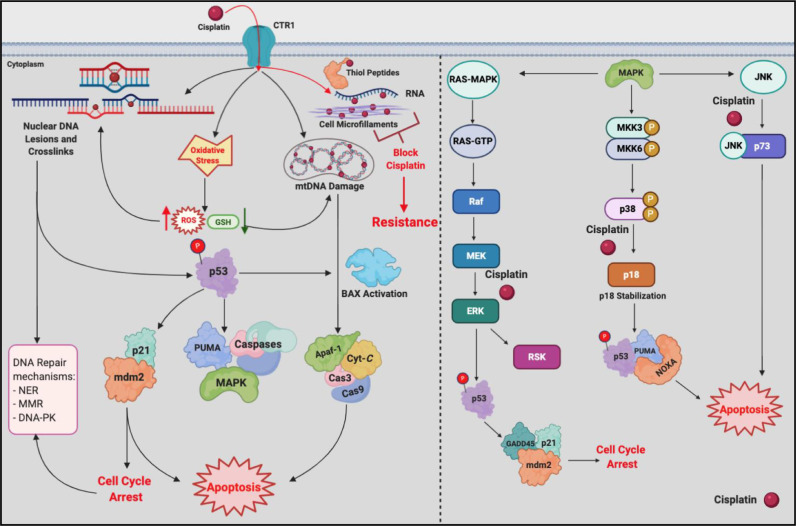
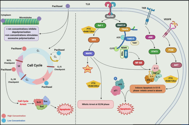
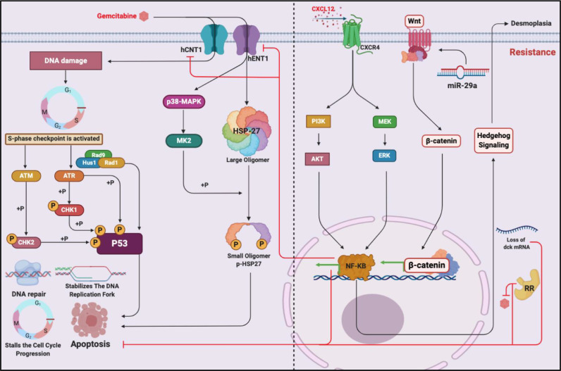
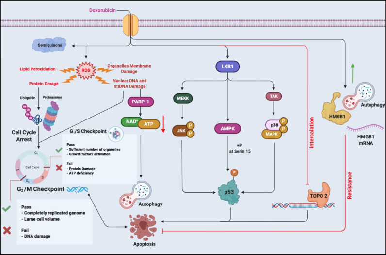
Chemotherapeutic drugs kill cancer cells or control their progression all over the patient's body, while radiation- and surgery-based treatments perform in a particular site. Based on their mechanisms of action, they are classified into different groups, including alkylating substrates, antimetabolite agents, anti-tumor antibiotics, inhibitors of topoisomerase I and II, mitotic inhibitors, and finally, corticosteroids. Although chemotherapeutic drugs have brought about more life expectancy, two major and severe complications during chemotherapy are chemoresistance and tumor relapse. Therefore, we aimed to review the underlying intracellular signaling pathways involved in cell death and resistance in different chemotherapeutic drug families to clarify the shortcomings in the conventional single chemotherapy applications. Moreover, we have summarized the current combination chemotherapy applications, including numerous combined-, and encapsulated-combined-chemotherapeutic drugs. We further discussed the possibilities and applications of precision medicine, machine learning, next-generation sequencing (NGS), and whole-exome sequencing (WES) in promoting cancer immunotherapies. Finally, some of the recent clinical trials concerning the application of immunotherapies and combination chemotherapies were included as well, in order to provide a practical perspective toward the future of therapies in cancer cases.

Keywords: Chemoresistance; Chemotherapeutic drugs; Combination chemotherapy; Death-related intracellular signaling; Intracellular signaling; Precision medicine; Resistance-related intracellular signaling.


Conflict of interest statement

Declaration of Competing Interest The authors declare no conflict of interest.

Figures

Image, graphical abstract
Graphical abstract
Fig. 1
Fig. 1
Platinum-based combinations in cisplatin constructs inter- and intrastrand adducts, namely lesions, within the nuclear and mitochondrial DNA. Besides it stimulates ROS and oxidative stress. Through the activation of p53, in which p21, Waf1, and MDM2 are recruited, and through the activation of Bax, in which the pro-caspase-9 and -3 are activated, the programed cell death, apoptosis pathways are ignited. To illustrate this, theses apoptosis pathways are accomplished through RAS-MAPK, p38-p18, and JNK cascades.
Fig. 2
Fig. 2
Paclitaxel targets microtubules and wreck the havoc on spindles in G2, prophase, and M phase. To be precise, it increases the polymerization of β-tubulin microtubules and prevents their depolymerization, which leads to the cell cycle arrest in G2/M phase. 10 nM of paclitaxel in 12 hrs stalls the cell cycle at the S phase, and by the administration of ≥9 nM ignites the apoptosis through the activation of Raf-1, whereas, administration of ≤9 nM recruits p53 and p21. TLR-4 is involved in two manners as well. Regarding apoptosis, JNK, NF-KB, MAPK, JAK, and STAT participate, where they lead the cell's fate to apoptosis through the dephosphorylation of Bad and Bax, and through the phosphorylation of Bcl2. Regarding the immune system, through the stimulation of TLR-4, paclitaxel recruits MyD88, MAPK, NF-KB which results in the expression and secretion of IL-1, IL-6, IL-2, IL-8, TNF-α, and IFN-γ.
Fig. 3
Fig. 3
Tyrosine kinase inhibitors (TKIs) are similar to ATP, hence they compete for ATP-binding domains in kinases of EGFRs. They inhibit PI3K/Akt, JAK/STAT, PLCγ, and Ras/Raf, therefore they are able to ignite apoptosis machinery in the absence of the aforementioned pathways. Gefitinib also increases the level of p27 and induces p38-MAPK as well which give rise to apoptosis. One major signaling that cells develop resistance against EGFR TKIs is through TGF-β signaling. Recruiting Smads 2,3, and 4, it stimulates cell survival and EMT. Moreover, Rab25 which has close interplay with integrin β1 correlates with TKI resistance. To be precise it phosphorylates Akt and ultimately assists survival and proliferation signaling cascades.
Fig. 4
Fig. 4
By the time gemcitabine is administered, it activates p38-MAPK that ignites apoptosis through MK2 and phosphorylation of HSP-27. Moreover, due to the DNA damage caused by gemcitabine, S phase checkpoint is activated, ATM/Chk2 and ATR/Chk1 are recruited which altogether with the company of 9-1-1-complex phosphorylate and activate p53. Regarding resistance, desmoplastic reaction seems to be accomplished through Hh signaling which accounts for poor drug delivery. RR also has been shown tot attenuate the activity of gemcitabine. Furthermore, CXCL12/CXCR4 stimulates PI3K/Akt, ERK, and NF-KB which along with Wnt/β catenin decreases the efficiency of gemcitabine therapy.
Fig. 5
Fig. 5
Doxorubicin forms adducts with Topoisomerase and DNA in a process, namely intercalation. When entered in to the target cell, DOX is oxidized into semiquinone which ROS are generated in its process. ROS further, causes lipid peroxidation, organelles’ membrane damage, DNA damage, and protein damage. Following the DNA damage PARP-1 is activated which leads cell toward either autophagy (providing that ATP and NAD+ resources are low), or G1/S arrest and apoptosis. Protein damages also lead to G2/M arrest which gives rise to apoptosis. Moreover, DOX can phosphorylate and activate p53 through JNK,AMPK, and p38-MAPK. However, increase in the protein or mRNA level of HMGB1 attenuates apoptosis and favors cell survival and resistance.
Fig. 6
Fig. 6
Etoposide, an anti-topoisomerase II agent, poisons the TopoIIcc and prevents the religation of DNA strands. Following the persistent DNA damage caused by etoposide, ATM/Chk2 are recruited that by the assist of Mre11/Rad50/NSB1 cause the S phase arrest which leads to apoptosis. Also through alternative signaling, namely NLK, ATM is phosphorylated which ultimately phosphorylates and activates p53 leading to p53. Nonetheless, SNP mutations like SNP 309 T/G forms a different subtype of MDM2 which inactivates p53 and accounts for etoposide resistance in some cases. Cyto C/Apaf-1/Cas-9 as well as Fas/FasL/Cas-8,10 are among other possible mechanisms described to play role in etoposide-cell death mechanisms.
Fig. 7
Fig. 7
MTOR is considered as the vital signaling cascade and as the crossroads within the realm of intracellular signaling owing to the fact that it serves different cellular functions under both physiological and pathological circumstances. It mainly acts through two complexes, namely mTORC1 and mTORC2. These complexes have a close interaction with upstream signaling cascades including, PI3K/Akt, Ras/ERK, and AMPK. The phosphorylation of Akt by the upstream signaling dampens the inhibitory function of TCS 2 which allows mTORC1 to perform its activities. The mTORC1 further regulates $E-BP1, S6K1, SREBP, STAT3, HIF-α, and PP2A in order to bring about lipid, protein, and nucleotide synthesis, as well as cell cycle progression and angiogenesis. The mTORC2, on the other hand, is modulated by mTORC1 through Grb-10 (in a feedback loop) and IRS-1 which ultimately favors cytoskeletal reorganization, cell movements, cell migration, and cell proliferation.

References

    1. Raimondi S., Maisonneuve P., Lowenfels A.B. Epidemiology of pancreatic cancer: an overview. Nat. Rev. Gastroenterol. Hepatol. 2009;6:699. - PubMed
    1. Midha S., Chawla S., Garg P.K. Modifiable and non-modifiable risk factors for pancreatic cancer: a review. Cancer Lett. 2016;381:269–277. - PubMed
    1. Anand P., Kunnumakara A.B., Sundaram C., Harikumar K.B., Tharakan S.T., Lai O.S., Sung B., Aggarwal B.B. Cancer is a preventable disease that requires major lifestyle changes. Pharm. Res. 2008;25:2097–2116. - PMC - PubMed
    1. Kushi L.H., Doyle C., McCullough M., Rock C.L., Demark-Wahnefried W., Bandera E.V., Gapstur S., Patel A.V., Andrews K., Gansler T. American cancer society guidelines on nutrition and physical activity for cancer prevention: reducing the risk of cancer with healthy food choices and physical activity. CA: Cancer J. Clin. 2012;62:30–67. - PubMed
    1. Mentella M.C., Scaldaferri F., Ricci C., Gasbarrini A., Miggiano G.A.D. Cancer and Mediterranean diet: a review. Nutrients. 2019;11:2059. - PMC - PubMed