. 2021 Dec;18(12):2278-2289.
doi: 10.1080/15476286.2021.1899653. Epub 2021 Mar 22.

Dihydrouridine synthesis in tRNAs is under reductive evolution in Mollicutes

Affiliations

Dihydrouridine synthesis in tRNAs is under reductive evolution in Mollicutes

Bruno Faivre et al. RNA Biol. 2021 Dec.

Abstract

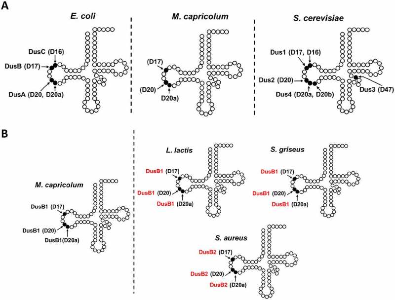
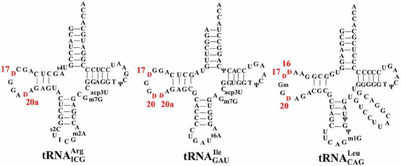
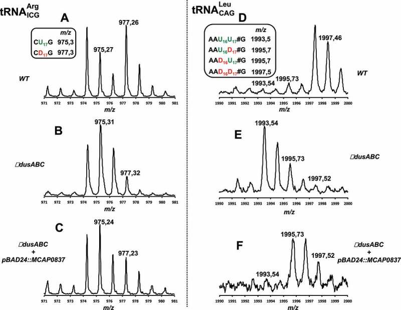
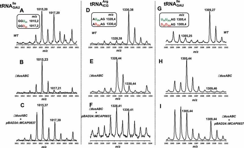
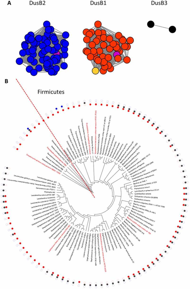
Dihydrouridine (D) is a tRNA-modified base conserved throughout all kingdoms of life and assuming an important structural role. The conserved dihydrouridine synthases (Dus) carries out D-synthesis. DusA, DusB and DusC are bacterial members, and their substrate specificity has been determined in Escherichia coli. DusA synthesizes D20/D20a while DusB and DusC are responsible for the synthesis of D17 and D16, respectively. Here, we characterize the function of the unique dus gene encoding a DusB detected in Mollicutes, which are bacteria that evolved from a common Firmicute ancestor via massive genome reduction. Using in vitro activity tests as well as in vivo E. coli complementation assays with the enzyme from Mycoplasma capricolum (DusBMCap), a model organism for the study of these parasitic bacteria, we show that, as expected for a DusB homolog, DusBMCap modifies U17 to D17 but also synthetizes D20/D20a combining therefore both E. coli DusA and DusB activities. Hence, this is the first case of a Dus enzyme able to modify up to three different sites as well as the first example of a tRNA-modifying enzyme that can modify bases present on the two opposite sides of an RNA-loop structure. Comparative analysis of the distribution of DusB homologs in Firmicutes revealed the existence of three DusB subgroups namely DusB1, DusB2 and DusB3. The first two subgroups were likely present in the Firmicute ancestor, and Mollicutes have retained DusB1 and lost DusB2. Altogether, our results suggest that the multisite specificity of the M. capricolum DusB enzyme could be an ancestral property.

Keywords: dihydrouridine; mollicutes; multisite-specificity; post-transcriptional modification; tRNA.


Conflict of interest statement

No potential conflict of interest was reported by the authors.

Figures

Figure 1.
Figure 1.
Location of D-sites in tRNA, site specificity of the known Dus enzymes and predicted site specificity of DusB corresponding enzymes in Firmicutes. (A) Schematic representation of the secondary structure of tRNA, showing the location of D residues and the corresponding Dus enzyme responsible for their synthesis in E. coli, eukaryotes and Mollicutes. (B) The presence of three D modifications in each gram+ bacteria presented are obtained by analysis of the tRNA sequences from Modomics. The specificity of DusB1 of Mycoplama capricolum has been validated here, experimentally. In red is indicated the predicted site specificity of the DusB homologue found in Lactococcus lactis, Streptomyces griseus and Staphylococcus aureus.
Figure 2.
Figure 2.
Absorbance spectrum and NADPH oxidase activity of DusBMCap . (A) In solid line (▬) is the spectrum of oxidized DusBMcap alone while the spectrum in dashed line (–) is upon denaturing condition in the presence of 0.1% SDS. Spectrum of a solution of free FMN is in dotted line (•••). (B) Steady state for the NADPH oxidase activity of DusBMcap. The open circles are the raw data while the solid line is the fit with a Michaelis Menten equation
Figure 3.
Figure 3.
In vitro dihydrouridine synthase activity of DusBMCap. Average of D/tRNA value of E. coli bulk tRNA in the presence of DusBMCap + NADPH incubated for 1 h at 30°C under anaerobic conditions and determined by the colorimetric assay described in the material and methods. A standard curve obtained with variable amounts of dihydrouracil was used to determine the concentration of D in tRNA. The error bars are calculated from five different sets of experiments. The upper panel shows the D-content of bulk E. coli tRNAs from (I) wild-type strain, (ii) ΔdusA strain, (ii) ΔdusA in the presence of recombinant DusBMcap and NADPH. The middle panel shows the D-content of bulk E. coli tRNAs from (i) wild-type strain, (ii) ΔdusB strain, (ii) ΔdusB in the presence of recombinant DusBMcap and NADPH. The lower panel is the D-content in bulk E. coli tRNAs from (i) wild-type strain, (ii) ΔdusC strain, (ii) ΔdusC in the presence of recombinant DusBMcap and NADPH
Figure 4.
Figure 4.
Secondary cloverleaf structure and sequence of E. coli tRNAICGArg, tRNAGAUIle and tRNACAGLeu. The positions of each D in the D-loop region are labelled in red
Figure 5.
Figure 5.
MALDI-TOF analysis of position 17 in tRNAs from E. coli ∆dusABC complemented with DusBMCap. (A), (B) and (C) are the MS relative isotope patterns of derived oligonucleotides after RNAse T1 treatment of tRNAICGArg isolated from wild type, ΔdusABC and ΔdusABC + pBAD24::MCAP0837, respectively. (D), (E) and (F) are the MS relative isotope patterns of derived oligonucleotides after RNAse T1 treatment of tRNACAGLeu isolated from wild type, ΔdusABC and ΔdusABC + pBAD24::MCAP0837, respectively. Summary of the tRNA-derived oligonucleotide fragments and their sizes (m/z) used for the identification of MCAP_0837 specificity are shown in the small boxes and are further detailed in Table S2
Figure 6.
Figure 6.
MALDI-TOF analysis of positions 20 and 20a in tRNAs from E. coli ∆dusABC complemented with DusBMCap.(A), (B) and (C) are the MS relative isotope patterns of derived oligonucleotides after RNAse A treatment of tRNAGAUIle isolated from wild type, ΔdusABC and ΔdusABC + pBAD24::MCAP0837, respectively. (D), (E) and (F) are the MS relative isotope patterns of derived oligonucleotides after RNAse T1 treatment of tRNAICGArg isolated from wild type, ΔdusABC and ΔdusABC + pBAD24::MCAP0837, respectively. (G), (H) and (I) are the MS relative isotope patterns of derived oligonucleotides after RNAse T1 treatment of tRNAGAUIle isolated from wild type, ΔdusABC and ΔdusABC + pBAD24::MCAP0837, respectively. Summary of the tRNA-derived oligonucleotide fragments and their sizes (m/z) used for the identification of MCAP_0837 specificity are shown in the small boxes and are further detailed in Table S2
Figure 7.
Figure 7.
Analysis of DusB subgroups in Firmicutes. (A) Results from the sequence similarity network analysis of 143 DusB proteins from Firmicutes performed on the EFI website using an alignment score threshold of 40 and visualized with Cytoscape, the DusB1 proteins are in red, the DusB2 proteins in blue, the DusB3 proteins in black, the M. capricolum DusB1 protein is in orange, the B. subtilis DusB1 and DusB2 proteins are in magenta; (B) Phylogenetic distribution analysis of the three DusB subgroups. Organisms discussed in the text are in red, the dotted line delineates the Firmicute radiation

Similar articles

Cited by

References

    1. Boccaletto P, Machnicka MA, Purta E, et al. MODOMICS: a database of RNA modification pathways. 2017 update. Nucleic Acids Res. 2018;46:D303–D7. - PMC - PubMed
    1. Suck D, Saenger W, Zechmeister K.. Conformation of the tRNA minor constituent dihydrouridine. FEBS Lett. 1971;12:257–259. - PubMed
    1. Dalluge JJ, Hashizume T, Sopchik AE, et al. Conformational flexibility in RNA: the role of dihydrouridine. Nucleic Acids Res. 1996;24:1073–1079. - PMC - PubMed
    1. Dyubankova N, Sochacka E, Kraszewska K, et al. Contribution of dihydrouridine in folding of the D-arm in tRNA. Org Biomol Chem. 2015;13:4960–4966. - PubMed
    1. Kim SH, Suddath FL, Quigley GJ, et al. Three-dimensional tertiary structure of yeast phenylalanine transfer RNA. Science. 1974;185:435–440. - PubMed

Publication types