. 2021 Mar 8;21(1):120.
doi: 10.1186/s12893-021-01134-1.

Taking a step down on the reconstruction ladder for head and neck reconstruction during the COVID-19 pandemic

Affiliations

Taking a step down on the reconstruction ladder for head and neck reconstruction during the COVID-19 pandemic

Haroon Ur Rashid et al. BMC Surg. .

Abstract

Background: Most of the head and neck cancers are time-critical and need urgent surgical treatment. Our unit is one of the departments in the region, at the forefront in treating head and neck cancers in Pakistan. We have continued treating these patients in the COVID-19 pandemic with certain modified protocols. The objective of this study is to share our experience and approach towards head and neck reconstruction during the COVID-19 pandemic.

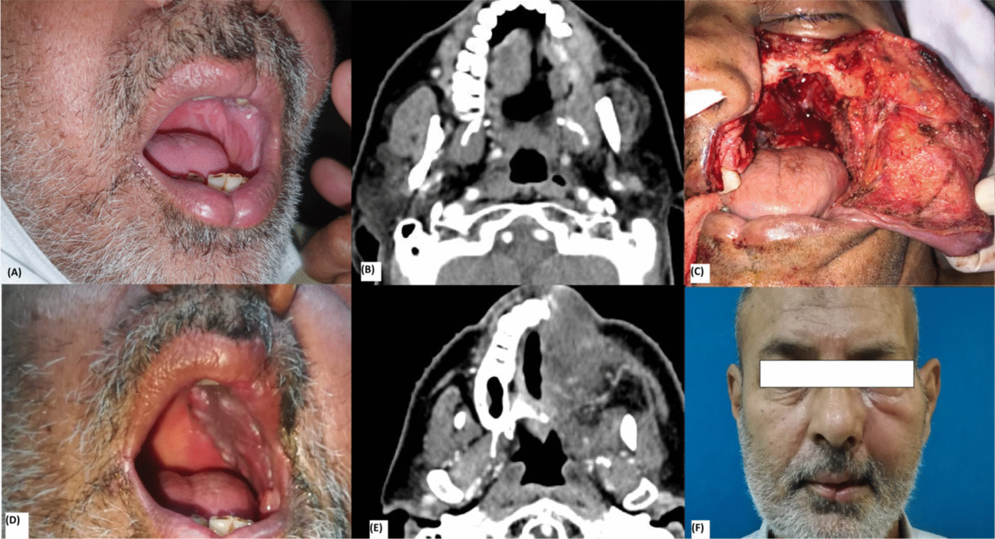
Results: There were a total of 31 patients, 20 (64.5%) were males and 11 (35.4%) patients were females. The mean age of patients was 52 years. Patients presented with different pathologies, i.e. Squamous cell carcinoma n = 26 (83.8%), mucoepidermoid carcinoma n = 2 (6.4%), adenoid cystic carcinoma n = 2 (6.4%) and mucormycosis n = 1 (3%). The reconstruction was done with loco-regional flaps like temporalis muscle flap n = 12 (38.7%), Pectoralis major myocutaneous flap n = 8 (25.8%), supraclavicular artery flap n = 10 (32.2%) and combination of fore-head, temporalis major and cheek rotation flaps n = 1 (3%). Defects involved different regions like maxilla n = 11 (35.4%), buccal mucosa n = 6 (19.3%), tongue with floor of mouth n = 6 (19.3%), mandible n = 4 (12.9%), parotid gland, mastoid n = 3 (9.6%) and combination of defects n = 1 (3%). Metal reconstruction plate was used in 3 (9.6%) patients with mandibular defects. All flaps survived, with the maximum follow-up of 8 months and minimum follow-up of 6 months.

Conclusion: Pedicled flaps are proving as the workhorse for head and neck reconstruction in unique global health crisis. Vigilant use of proper PPE and adherence to the ethical principles proves to be the only shield that will benefit patients, HCW and health system.

Keywords: Head and neck reconstruction; Pedicled flaps in free flap era; Reconstruction in COVID 19 pandemic.


Conflict of interest statement

There are no competing interests.

Figures

Fig. 1
Fig. 1
Case 1 a Young lady with large defect right cheek, nasal dorsum and palate with visible tongue at base. b Markings for forehead flap, temporalis muscle flap and cheek rotation advancement flaps. c Temporalis muscle flap inset done to reconstruct palate and fill the dead space. d Per-operative pic showing inset of forehead and cheek flaps. e Intra-oral view showing good mucosalization of temporalis muscle. f Frontal view of face at early follow up period
Fig. 2
Fig. 2
Case 2 a Case 2 Young male with recurrent Squamous cell carcinoma lower lip and mandible. There is a visible supraclavicular flap of previous surgery. b CT scan showing extent of tumor to bone and floor of mouth. c Per-operative view of wide local excision showing soft tissue and boney defect. d Per-operative view showing pectoralis major myocutaneous flap after inset and Karapandzic technique used for lip defect. e Early followup picture. f Frontal view of patient showing nicely healed wounds
Fig. 3
Fig. 3
Case 3 a Middle age male with large fungating mass of the left maxilla. b CT scan showing tumor invading maxillary bone and extending into palate. c Intra-operative picture showing large defect after wide local excision. d Early followup picture showing temporalis muscle at left palatal half. e Picture showing flap mucosalization at 4 weeks follow up. f Late follow up picture with good facial contours with mild temporal hollowing
Fig. 4
Fig. 4
Case 4 a Middle aged male with biopsy proven well differentiated squamous cell carcinoma of the left maxilla. b CT scan with contrast showing enhancement in the left maxilla sinus, left sided palate with destruction of the left maxillary arch. c Picture showing a moderate volume with large surface area defect after wide local excision of the tumor (subtotal maxillectomy) including resection of maxillary arch, palate and anterior and lateral walls with preservation of the orbital floor. d Four weeks follow up picture showing good mucosalisation of the temporal muscle flap. e Follow up CT scan of the patient showing temporalis muscle flap filling the defect. f Late follow up picture, patient has good facial contours with mild temporal hollowing
Fig. 5
Fig. 5
Case 5 a A young female patient presented with an ulcerative lesion of the right buccal mucosa, Biopsy reported well differentiated squamous cell carcinoma. b After resection of the tumor, An extended supraclavicular flap was elevated. c De-epithelialization of the proximal part of the flap was done, which was tunneled under the neck skin into the defect. d picture showing the flap inset into the defect and primary closure of the donor site. e, f 2 weeks follow up of the patient showing right sided cheek edema with good flap mucosalisation and good healing of the donor area
Fig. 6
Fig. 6
Case 6 a Patient with squamous cell carcinoma of body of the right mandible with left mandible extension with involvement of the floor of the mouth (T4a) b He had limited mouth opening with restricted tongue movements c Orthopantomogram (OPG) showing cortical destruction around the mandibular symphysis. d Enbloc Resected specimen showing mandibular arch with part of the floor of the mouth and bilateral neck nodes. e right lateral Xray view showing the reconstructed lower jaw with metallic reconstruction plate. f soft tissue coverage was provided with myocutaneous pectoralis major flap. Picture showing healed donor site
Fig. 7
Fig. 7
Case 7 a A young female patient presented with diagnosed case of adenoid cystic carcinoma of the right parotid with involvement of the middle ear b lateral view showing the extension of the tumor into the mastoid area. c Marking of the resection with 1 cm margins. d Excision involved peri-auricular skin, superficial parotidectomy, external and internal auditory meatus. Ear was intact by a bridge of skin at the root of the helix. e A supraclavicular artery flap was designed according to the defect size f Picture showing flap inset and donor site closure over a Redivac drain g Lateral view of patient at 1 month follow up
Fig. 8
Fig. 8
Shows follow-up pictures of same patients at 6 months

References

    1. World Health Organization. Coronavirus disease 2019 (COVID-2019) Situation Report-51. https://www.who.int/docs/default-source/coronaviruse/situation-reports/2....
    1. Coronavirus Disease 2019: For Healthcare Professionals. https://www.cdc.gov/coronavirus/2019-nCoV/hcp/index.html.
    1. CMS Adult Elective Surgery and Procedures Recommendations. https://cms.gov/files/document/covid-elective-surgery-recommendations.pdf.
    1. Wong CH, Wei FC. Microsurgical free flap in head and neck reconstruction. Head Neck. 2010;32(9):1236–1245. doi: 10.1002/hed.21284. - DOI - PubMed
    1. Chie WC, Hong RL, Lai CC, Ting LL, Hsu MM. Quality of life in patients of nasopharyngeal carcinoma: validation of the Taiwan Chinese version of the EORTC QLQ-C30 and the EORTC QLQ-H&N35. Qual Life Res. 2003;12(1):93–98. doi: 10.1023/A:1022070220328. - DOI - PubMed