. 2021 Mar 9;8(1):18.
doi: 10.1186/s40779-021-00308-5.

Recombinant human epidermal growth factor combined with vacuum sealing drainage for wound healing in Bama pigs

Affiliations

Recombinant human epidermal growth factor combined with vacuum sealing drainage for wound healing in Bama pigs

Shuai Wei et al. Mil Med Res. .

Abstract

Background: Vacuum sealing drainage (VSD) and epidermal growth factor (EGF) both play an important role in the treatment of wounds. This study aims to explore the effects of the combination of VSD and EGF on wound healing and the optimal concentration and time of EGF.

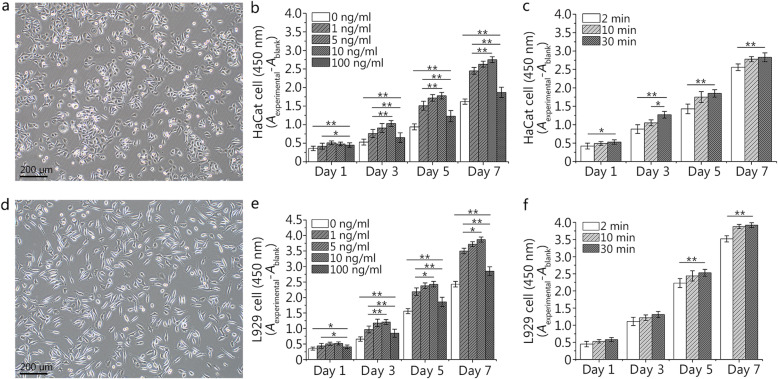
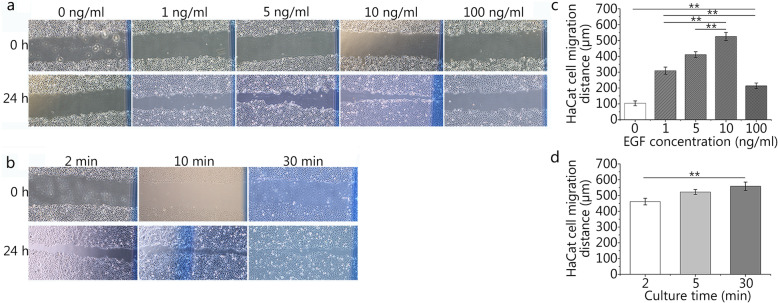
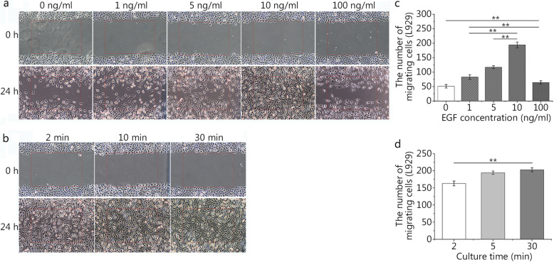
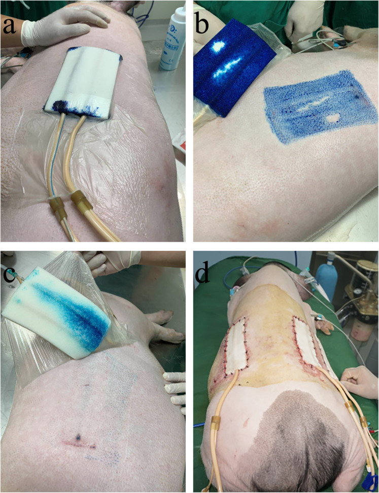
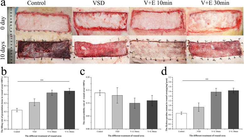
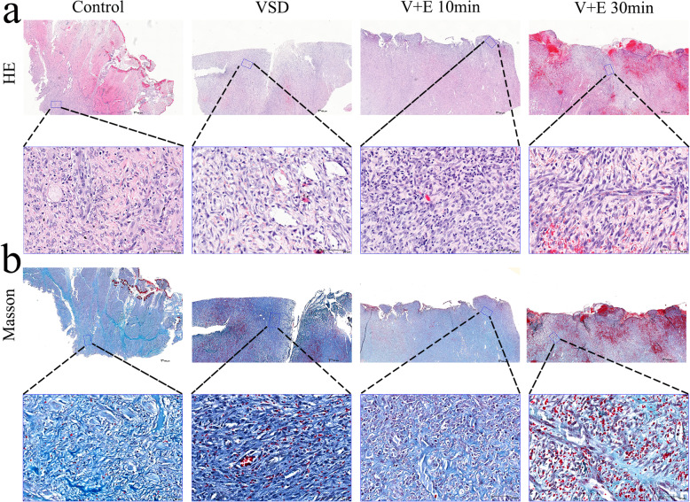
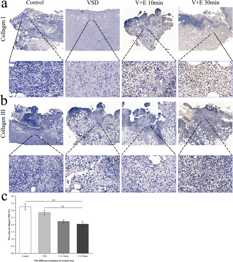
Methods: We tested the proliferation and migration capacity of HaCaT and L929 cells at different EGF concentrations (0, 1, 5, 10, and 100 ng/ml) and different EGF action times (2, 10, and 30 min). A full-thickness skin defect model was established using male, 30-week-old Bama pigs. The experiment included groups as follows: routine dressing change after covering with sterile auxiliary material (Control), continuous negative pressure drainage of the wound (VSD), continuous negative pressure drainage of the wound and injection of EGF 10 min followed by removal by continuous lavage (V + E 10 min), and continuous negative pressure drainage of the wound and injection of EGF 30 min followed by removal by continuous lavage (V + E 30 min). The wound healing rate, histological repair effect and collagen deposition were compared among the four groups.

Results: An EGF concentration of 10 ng/ml and an action time of 10 min had optimal effects on the proliferation and migration capacities of HaCaT and L929 cells. The drug dispersion effect was better than drug infusion after bolus injection effect, and the contact surface was wider. Compared with other groups, the V + E 10 min group promoted wound healing to the greatest extent and obtained the best histological score.

Conclusions: A recombinant human epidermal growth factor (rhEGF) concentration of 10 ng/ml can promote the proliferation and migration of epithelial cells and fibroblasts to the greatest extent in vitro. VSD combined with rhEGF kept in place for 10 min and then washed, can promote wound healing better than the other treatments in vivo.

Keywords: Epidermal growth factor; Full-thickness skin defect; Skin wound healing; Vacuum sealing drainage.


Conflict of interest statement

The authors declare that they have no competing interests.

Figures

Fig. 1
Fig. 1
Effects of different concentrations and durations of epidermal growth factor (EGF) on the proliferation of HaCaT and L929 cells. a. Observation of HaCaT cells at day 2 under an inverted microscope. b. The effects of different concentrations (0, 1, 5, 10, and 100 ng/ml) of EGF on the proliferation of HaCaT cells at different time. c. The effects of different duration (2, 10, and 30 min) and a set concentration (10 ng/ml) of EGF on the proliferation of HaCaT cells at different time. d. Observation of L929 cells at day 2 under an inverted microscope. e. The effects of different concentrations (0, 1, 5, 10, and 100 ng/ml) of EGF on the proliferation of L929 cells at different time. f. The effects of different duration (2, 10, and 30 min) and a set concentration (10 ng/ml) of EGF on the proliferation of L929 cells at different time. Data are shown as means ± SD, *P < 0.05, **P < 0.01
Fig. 2
Fig. 2
Effects of different concentrations and durations of epidermal growth factor (EGF) on HaCaT cell migration. a and c. The effects of different concentrations (0, 1, 5, 10, and 100 ng/ml) of EGF on the migration distance of HaCaT cells (0 ng/ml: 103.37 ± 15.37 μm, 1 ng/ml: 309.34 ± 22.68 μm, 5 ng/ml: 410.63 ± 18.60 μm, 10 ng/ml: 525.24 ± 25.79 μm, 100 ng/ml: 213.94 ± 17.40 μm). b and d. The effects of different durations (2, 10, and 30 min) and a set concentration (10 ng/ml) of EGF on the migration distance of HaCaT cells (2 min: 461.46 ± 20.84 μm, 10 min: 522.18 ± 15.59 μm, 30 min: 558.35 ± 26.68 μm). The scale bar is 500 μm. Data are shown as means ± SD, **P < 0.01
Fig. 3
Fig. 3
Effects of different concentrations and durations of epidermal growth factor (EGF) on L929 cell migration. a and c. The effects of different concentrations (0, 1, 5, 10, and 100 ng/ml) of EGF on the migration number of L929 cells (0 ng/ml: 51 ± 6, 1 ng/ml: 83 ± 8, 5 ng/ml: 117 ± 5, 10 ng/ml: 194 ± 10, 100 ng/ml: 64 ± 6). b and d. The effects of different duration (2, 10, and 30 min) and a set concentration (10 ng/ml) of EGF on the migration number of L929 cells (2 min: 163 ± 7, 10 min: 194 ± 5, 30 min: 203 ± 6). The scale bar is 500 μm. Data are shown as means ± SD, **P < 0.01
Fig. 4
Fig. 4
Effects of different administration methods on drug dispersion during vacuum sealing drainage (VSD) treatment and a trauma model in Bama pigs. a. Image of the opening the VSD negative pressure device for suction after injection with a 15-ml syringe. b. Image of the dispersion effect after injection with the 15-ml syringe. c. Image of the dispersion effect after injection with an infusion set. d. The trauma model of the Bama pig after surgical operation using VSD and epidermal growth factor (EGF)
Fig. 5
Fig. 5
General view of the postoperative wound and evaluation of the effect of epidermal growth factor (EGF) combined with vacuum sealing drainage (VSD) in the treatment of wounds. a. General views of the postoperative wound and the wound 10 d after the operation. b. The filling rate of granulation tissue in the wound area 10 d after the operation (Control: 0.22 ± 0.02, VSD: 0.31 ± 0.04, V + E 10 min: 0.42 ± 0.03, V + E 30 min: 0.44 ± 0.03). c. The contractibility rate of the wound area 10 d after the operation (Control: 0.14 ± 0.01, VSD: 0.13 ± 0.03, V + E 10 min: 0.10 ± 0.02, V + E 30 min: 0.11 ± 0.02). d. The hydroxyproline content in the wound area 10 d after the operation (Control: 0.67 ± 0.05 μg/mg*prot, VSD: 0.93 ± 0.18 μg/mg*prot, V + E 10 min: 1.58 ± 0.16 μg/mg*prot, V + E 30 min: 1.64 ± 0.10 μg/mg*prot). Data are shown as means ± SD, **P < 0.01
Fig. 6
Fig. 6
Histological evaluation of epidermal growth factor (EGF) combined with vacuum sealing drainage (VSD) in the treatment of wounds. a. HE staining of the wound 10 d after operation; the following picture is an enlargement of the local area from the above picture. b. Masson staining of the wound 10 d after the operation; the following picture is the enlargement of the local area from the above picture. The scale bar in the global image above is 500 μm, and the scale bar in the enlarged image below is 70 μm
Fig. 7
Fig. 7
Immunohistochemical evaluation of epidermal growth factor (EGF) combined with vacuum sealing drainage (VSD) in the treatment of wounds. a. Immunohistochemical staining of collagen I in the wound 10 d after the operation; the following picture is an enlargement of the local area from the above picture. b. Immunohistochemical staining of collagen III in the wound 10 d after the operation; the following picture is an enlargement of the local area from the above picture. c. The ratio of collagen I/III in the wound area 10 d after the operation (Control: 3.26 ± 0.22, VSD: 2.85 ± 0.17, V + E 10 min: 2.24 ± 0.12, V + E 30 min: 2.05 ± 0.17). The scale bar in the global image above is 500 μm, and the scale bar in the enlarged image below is 70 μm. Data are shown as means ± SD, **P < 0.01

References

    1. Jiang B, Liang S, Peng ZR, Cong H, Levy M, Cheng Q, et al. Transport and public health in China: the road to a healthy future. Lancet. 2017;390(10104):1781–1791. doi: 10.1016/S0140-6736(17)31958-X. - DOI - PMC - PubMed
    1. Mam L, Kenter SB, Au FL, Wjm VG, Middelkoop E, Bos JD. Fibroblasts derived from chronic diabetic ulcers differ in their response to stimulation with EGF, IGF-I, bFGF and PDGF-AB compared to controls. Eur J Cell Biol. 2002;81(3):153–160. doi: 10.1078/0171-9335-00228. - DOI - PubMed
    1. Singer AJ, Clark RA. Cutaneous wound healing. N Engl J Med. 1999;341(10):738–746. doi: 10.1056/NEJM199909023411006. - DOI - PubMed
    1. Xu P, Wu Y, Zhou L, Yang Z, Zhang X, Hu X, et al. Platelet-rich plasma accelerates skin wound healing by promoting re-epithelialization. Burns Trauma. 2020;8:tkaa028. doi: 10.1093/burnst/tkaa028. - DOI - PMC - PubMed
    1. Greenhalgh DG. The role of growth factors in wound healing. J Trauma. 1996;41(1):159–167. doi: 10.1097/00005373-199607000-00029. - DOI - PubMed

Publication types

MeSH terms

Substances

LinkOut - more resources