. 2021 Mar 8;4(1):300.
doi: 10.1038/s42003-021-01835-6.

A stalled-ribosome rescue factor Pth3 is required for mitochondrial translation against antibiotics in Saccharomyces cerevisiae

Affiliations

A stalled-ribosome rescue factor Pth3 is required for mitochondrial translation against antibiotics in Saccharomyces cerevisiae

Soichiro Hoshino et al. Commun Biol. .

Abstract

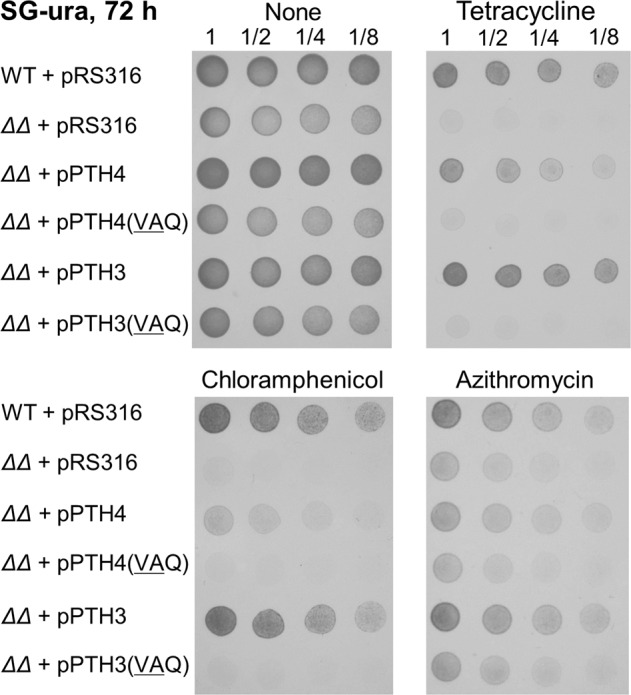
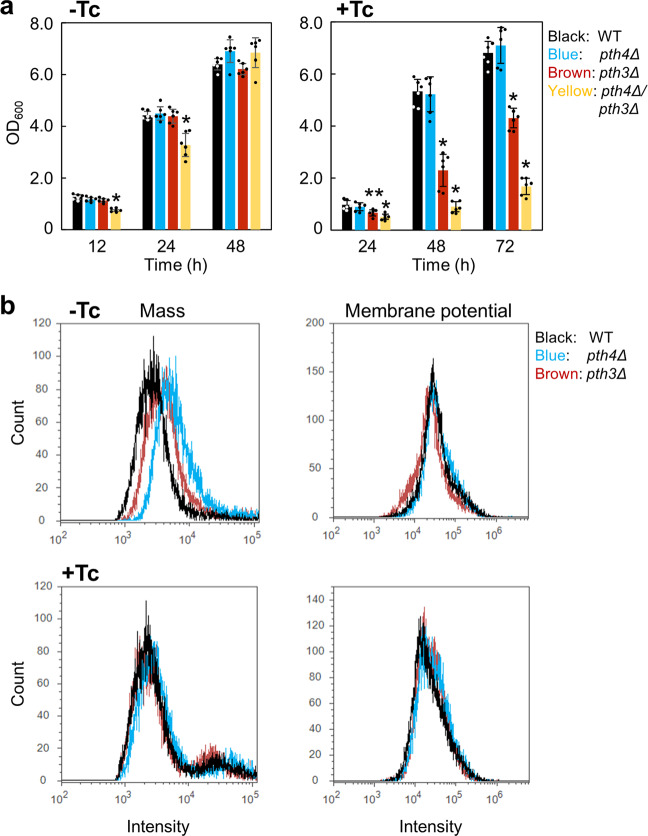
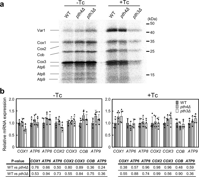
Mitochondrial translation appears to involve two stalled-ribosome rescue factors (srRFs). One srRF is an ICT1 protein from humans that rescues a "non-stop" type of mitochondrial ribosomes (mitoribosomes) stalled on mRNA lacking a stop codon, while the other, C12orf65, reportedly has functions that overlap with those of ICT1; however, its primary role remains unclear. We herein demonstrated that the Saccharomyces cerevisiae homolog of C12orf65, Pth3 (Rso55), preferentially rescued antibiotic-dependent stalled mitoribosomes, which appear to represent a "no-go" type of ribosomes stalled on intact mRNA. On media containing a non-fermentable carbon source, which requires mitochondrial gene expression, respiratory growth was impaired significantly more by the deletion of PTH3 than that of the ICT1 homolog PTH4 in the presence of antibiotics that inhibit mitochondrial translation, such as tetracyclines and macrolides. Additionally, the in organello labeling of mitochondrial translation products and quantification of mRNA levels by quantitative RT-PCR suggested that in the presence of tetracycline, the deletion of PTH3, but not PTH4, reduced the protein expression of all eight mtDNA-encoded genes at the post-transcriptional or translational level. These results indicate that Pth3 can function as a mitochondrial srRF specific for ribosomes stalled by antibiotics and plays a role in antibiotic resistance in fungi.


Conflict of interest statement

The authors declare no competing interests.

Figures

Fig. 1
Fig. 1. Susceptibility of PTH4- and/or PTH3-deficient mutants of S. cerevisiae to temperature and antibiotics.
a The wild type (WT), pth4Δ, pth3Δ, and pth4∆pth3∆ were serially diluted and then spotted onto YPD and YPG plates containing glucose and glycerol as a carbon source, respectively. Plates were incubated at 30 °C or 37 °C for the indicated hours. The results of all of the spotting assays in the present study are shown for one experiment representative of at least three independent experiments. b The four strains were serially diluted and then spotted onto YPG plates containing the following antibiotics, with the concentration shown in parentheses (µg/mL): tetracycline (100), oxytetracycline (200), doxycycline (100), azithromycin (75), erythromycin (20), chloramphenicol (1000), paromomycin (100), tobramycin (100), and streptomycin (1000). Plates were incubated at 30 °C for 48 h (tetracycline, oxytetracycline, and doxycycline) or 72 h (the others). Corresponding images of cells grown on YPD plates for 24 h are shown in Supplementary Fig. 2.
Fig. 2
Fig. 2. Suppression of the antibiotic susceptibility phenotype of the double-gene deletion mutant by plasmid-borne PTH3 or PTH4.
The pth4∆pth3∆ mutant (represented by ∆∆) was transformed with the plasmid pRS316 harboring a genomic region including PTH4 and PTH3, termed pPTH4 and pPTH3, and the resultant strains were labeled ∆∆+pPTH4 and ∆∆+pPTH3, respectively. Each strain was diluted and spotted onto SG-ura plates containing no antibiotic, 100 µg/mL tetracycline, 1000 µg/mL chloramphenicol or 75 µg/mL azithromycin. As a control, pth4∆pth3∆ as well as the wild type was transformed with an empty plasmid, pRS316. In addition, pth4∆pth3∆ was transformed with the plasmid in which the GGQ motif residues were changed to VAQ in pPTH4 and pPTH3, termed pPTH4(VAQ) and pPTH3(VAQ), respectively. Corresponding images of cells grown on SC-ura plates for 12 h are shown in Supplementary Fig. 4a.
Fig. 3
Fig. 3. Effects of the deletion of PTH4 or PTH3 on mitochondrial properties.
a The three mutants and the wild type were inoculated into liquid YPG media in the absence or presence of 500 µg/mL tetracycline (Tc) with a starting OD600 of 0.01. Growth was monitored by measuring the OD600 values at the indicated time points at 30 °C. Data are shown as the mean ± standard deviation of six independent experiments. Asterisks indicate significant difference from the wild type (Student’s t test, *P < 0.001 and **P < 0.01). b Mitochondrial mass (left) and membrane potential (right) in the wild type (WT), pth4Δ, pth3Δ, and pth4∆pth3∆ grown in liquid YPG media for 48 h were measured at room temperature (about 25 °C) by FCM using MitoTracker Green FM and MitoTracker Red CMXRos, respectively, in the absence or presence of 500 µg/mL tetracycline (Tc).
Fig. 4
Fig. 4. Effects of the deletion of PTH4 or PTH3 on mitochondrial translation and transcription in the absence or presence of tetracycline.
a Mitochondrial translation products synthesized in organelles from the wild type (WT), pth4Δ, and pth3Δ grown in YPG media for 24 h in the absence or presence of 500 µg/mL tetracycline (Tc). Fresh mitochondria isolated from each strain were incubated with [35S]methionine in translational buffer. Mitochondrial lysates were analyzed by 16% SDS-PAGE and autoradiography. The corresponding Coomassie blue-stained gels are shown in Supplementary Fig. 7a. Quantitative comparison of mitochondrial translation products is shown in Supplementary Fig. 7b. b qPCR data of all mtDNA protein-coding genes, except for VAR1, from total RNAs extracted from the three strains in the absence or presence of 500 µg/mL Tc. Data are shown as the mean ± standard deviation of five independent experiments. P values are shown in the tables. There were no statistical significant differences between the wild type and each mutant in the absence or presence of Tc (Student’s t test, p > 0.1). The mRNA expression of VAR1 was described in the text.
Fig. 5
Fig. 5. PTH activities of Pth4 and Pth3 toward non-stop or no-go ribosomes using the E. coli-based reconstituted translation system.
a In vitro translation of the non-stop template with recombinant His-tagged Pth4 and Pth3 proteins. Each recombinant protein was added to the solution in which a 15-min in vitro translation reaction had been performed using the non-stop templates. The resulting mixture, incubated for 5 min, was analyzed by NuPAGE, shown in the left panel. Each gel was visualized using a laser-based fluorescent gel scanner. Released peptides and peptidyl-tRNA indicate Crp proteins and Crp-tRNA, respectively. The final concentration of each recombinant protein is shown in each lane. An asterisk indicates a background band (≈30 kDa) that appears without the addition of template DNA or mRNA, as previously shown in the manufacturer’s technical note. The right panel shows the extent of peptidyl-tRNA hydrolysis (relative PTH activity) given as the ratio of the band intensity of released peptides to that of peptidyl-tRNA plus that of released peptides at the protein concentrations indicated. Data are shown as the mean ± standard deviation of three independent experiments. b Comparison of concentrations between Pth4 and Pth3 proteins required for similar PTH activities toward non-stop or no-go ribosomes. Each upper panels show a schematic drawing of a ribosome stalled on non-stop or no-go mRNA. The lower panels show that ≈60% PTH activities for non-stop and no-go ribosomes require the indicated concentrations of Pth4 or Pth3. Data are shown as the mean ± standard deviation of five independent experiments. An example of gel images is shown in Supplementary Fig. 8e.

Similar articles

Cited by

References

    1. Moore SD, Sauer RT. Ribosome rescue: tmRNA tagging activity and capacity in Escherichia coli. Mol. Microbiol. 2005;58:456–466. doi: 10.1111/j.1365-2958.2005.04832.x. - DOI - PubMed
    1. Himeno H, Nameki N, Kurita D, Muto A, Abo T. Ribosome rescue systems in bacteria. Biochimie. 2015;114:102–112. doi: 10.1016/j.biochi.2014.11.014. - DOI - PubMed
    1. Keiler KC. Mechanisms of ribosome rescue in bacteria. Nat. Rev. Microbiol. 2015;13:285–297. doi: 10.1038/nrmicro3438. - DOI - PubMed
    1. Janssen BD, Hayes CS. The tmRNA ribosome-rescue system. Adv. Protein Chem. Struct. Biol. 2012;86:151–191. doi: 10.1016/B978-0-12-386497-0.00005-0. - DOI - PMC - PubMed
    1. Huter P, Muller C, Arenz S, Beckert B, Wilson DN. Structural basis for ribosome rescue in bacteria. Trends Biochem. Sci. 2017;42:669–680. doi: 10.1016/j.tibs.2017.05.009. - DOI - PubMed

Publication types

MeSH terms