. 2021 Mar 8;11(1):5418.
doi: 10.1038/s41598-021-84123-x.

3D-microtissue derived secretome as a cell-free approach for enhanced mineralization of scaffolds in the chorioallantoic membrane model

Affiliations

3D-microtissue derived secretome as a cell-free approach for enhanced mineralization of scaffolds in the chorioallantoic membrane model

Lukas Otto et al. Sci Rep. .

Abstract

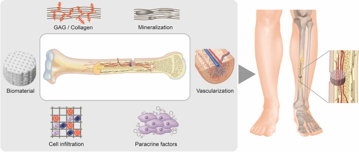
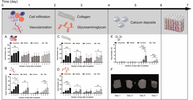
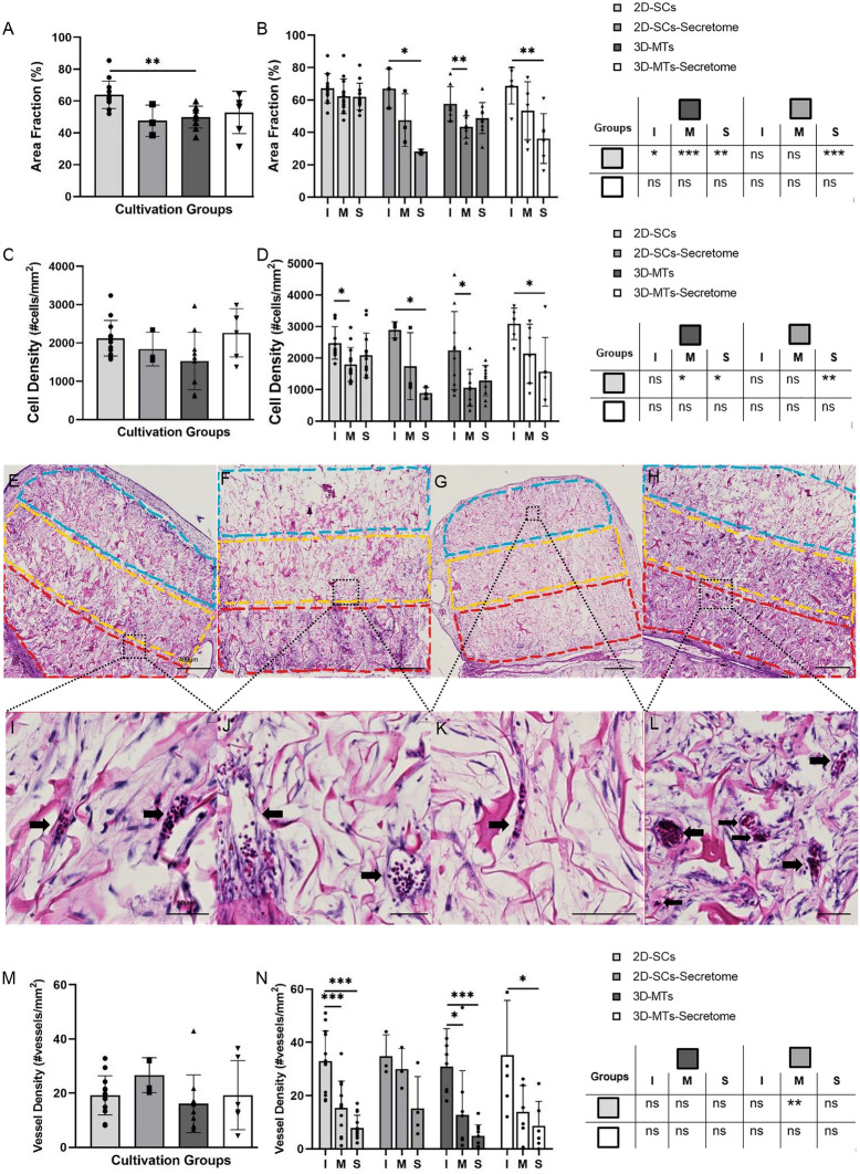
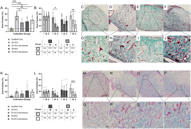
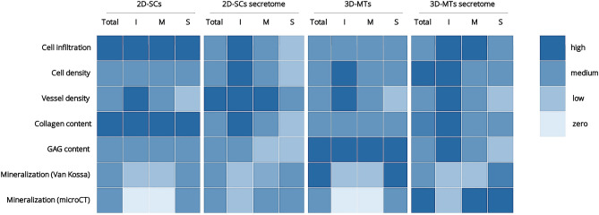
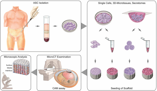
Bone regeneration is a complex process and the clinical translation of tissue engineered constructs (TECs) remains a challenge. The combination of biomaterials and mesenchymal stem cells (MSCs) may enhance the healing process through paracrine effects. Here, we investigated the influence of cell format in combination with a collagen scaffold on key factors in bone healing process, such as mineralization, cell infiltration, vascularization, and ECM production. MSCs as single cells (2D-SCs), assembled into microtissues (3D-MTs) or their corresponding secretomes were combined with a collagen scaffold and incubated on the chicken embryo chorioallantoic membrane (CAM) for 7 days. A comprehensive quantitative analysis was performed on a cellular level by histology and by microcomputed tomography (microCT). In all experimental groups, accumulation of collagen and glycosaminoglycan within the scaffold was observed over time. A pronounced cell infiltration and vascularization from the interface to the surface region of the CAM was detected. The 3D-MT secretome showed a significant mineralization of the biomaterial using microCT compared to all other conditions. Furthermore, it revealed a homogeneous distribution pattern of mineralization deposits in contrast to the cell-based scaffolds, where mineralization was only at the surface. Therefore, the secretome of MSCs assembled into 3D-MTs may represent an interesting therapeutic strategy for a next-generation bone healing concept.


Conflict of interest statement

The authors declare no competing interests.

Figures

Figure 1
Figure 1
Key factors for bone regeneration in the applications of bone grafts in combination with stem cells or their secreted factors. In addition to calcification, components of the extracellular matrix such as glycosaminoglycan (GAG) and collagen play an important role in the healing process. For the functionality of the bone and the viability of the graft, the enabling of cell infiltration and thus vascularization of the construct are also important prerequisites. The selection of an appropriate biomaterial as scaffold is of fundamental relevance, as this must be able the interaction of the key factors mentioned above. When combined with stem cells or their secretomes only, the regeneration is further promoted by paracrine factors.
Figure 2
Figure 2
Time course regarding the occurrence and impact of key factors on the calcification process analyzed in collagen scaffold. The timing of the various factors involved was investigated (AF) using the CAM assay. A marked increase in cell infiltration and vessel density in the scaffold was observed on day 1 (A, B). On day 3, there was a distinct increase in collagen and glycosaminoglycan (GAG) content (C, D) and from day 5 on, an accumulation of calcium deposits could be sufficiently detected (E), which could also be shown by microCT analysis (F). Schematic scaffold with the localization of key factors is shown after 7 days of incubation on the CAM.
Figure 3
Figure 3
Histological analysis of cell infiltration (%) and cell density (cells/mm2) into the natural collagen scaffold (Optimaix). Histological analysis of the infiltration (A, B), cell density (C, D) and vessel density (M, N) of the four cultivation groups are assessed on day 7. Results are presented in a group comparison of the total scaffold (A, C, M) and more specified in differing three ROIs (interface (I = red), middle (M = yellow) and the surface (S = blue)) shown in (B, D, N). Significances of intra-group calculations are demonstrated within the graph whereas inter-group differences with its corresponding ROIs are listed on the separate table next to the graph (B, D, N). The results are illustrated with an exemplary out-take of each experimental group using single cells (E, I), 2D-SCs secretome (F, J), 3D-microtissues (G, K), 3D-MT secretome (H, L). The scale bar was defined with 400 µm in the overview examples (EH) and 50 µm for the more detailed examples (IL). Statistical results were established with a one-way ANOVA considering only one independent factor and a two-way ANOVA considering two categorical factors and the effect of the categorical factors on each other. P-values were considered significant by the APA-System: 0.12 (ns), 0.033 (*), 0.002 (**), < 0.001 (***).
Figure 4
Figure 4
Histological analysis of collagen and glycosaminoglycan formation into the natural collagen scaffold (Optimaix). Histological analysis of collagen (AJ) and GAG (KT) of the four cultivation groups are assessed on day 7. An additional scaffold only group was assessed to provide a reference what amount is stained by the scaffold itself. Results are presented in a group comparison of the total scaffold (A, K) and more specified in differing three ROIs (interface (I), middle (M) and the surface (S)) (B, L). Significances of intra-group calculations are demonstrated within the graph whereas inter-group differences with its corresponding ROIs are shown on the separate tables. The results are illustrated with an exemplary out-take of each cultivation group using single-cells (C, G, M, Q), 2D-SCs-secretome (D, H, N, R), 3D-microtissues (E, I, O, S), 3D-MT-Secretome (F, J, P, T). An exemplary part of the interface region is shown for each group (GJ, QT). The scale bar was defined with 400 µm in the overview examples (CF, MP) in both stains and 100 µm for the more detailed examples of collagen formation (GJ) and 50 µm of GAG formation (QT). Statistical results were established with a one-way ANOVA considering only one independent factor and a two-way ANOVA considering two categorical factors and the effect of the categorical factors on each other. P-values were considered significant by the APA-System: 0.12 (ns), 0.033 (*), 0.002 (**), < 0.001 (***).
Figure 5
Figure 5
Histological and microCT analysis of the calcification potential of ASCs in the natural collagen scaffold (Optimaix). Calcification was assessed with Von Kossa histological analysis (AJ) and a microCT analysis (KO). Results are presented in a group comparison of the total scaffold (A, K) and more specified in differing three ROIs (interface (I), middle (M) and the surface (S); shown in B). Significance of intra-group calculations are demonstrated within the graph whereas inter-group differences with its corresponding ROIs are shown on the separate table, bellow the graph (B). The results are illustrated with an exemplary out-take of each cultivation group using single-cells (C, G, L, Q), 2D-SCs secretome (D, H, M, R), 3D-microtissues (E, I, N, S), 3D-MT secretome (F, J, O, T). An exemplary part of the surface region is shown for each group (GJ). Two scaffolds were placed as onplants on the CAM of each chicken egg (P). View of the scaffolds from the top shows a tendency of calcification (QT). Dotted line marks edge of scaffolds. The scale bar was defined with 400 µm in the overview examples (CF) and 50 µm for the more detailed examples (GJ). Statistical results were established with a one-way ANOVA considering only one independent factor and a two-way ANOVA considering two categorical factors and the effect of the categorical factors on each other. P-values were considered significant by the APA-System: 0.12 (ns), 0.033 (*), 0.002 (**), < 0.001 (***).
Figure 6
Figure 6
Heat map showing overview of key results. All quantitative results were assigned to one of the following categories zero, low, medium and high and color-coded accordingly. The investigated scaffolds of the four experimental groups 2D-single cells (2D-SCs), 3D-microtissues (3D-MTs) and corresponding secretomes were either analyzed as an entire scaffold (Total) or one of the three regions interface (I), middle (M) and surface (S).
Figure 7
Figure 7
Schematic experimental setup for the investigation of calcification capacity of Optimaix scaffold in combination with human adipose tissue-derived mesenchymal stem cells or secretome. After isolation of human ASCs, the cells are cultured either as 2D -single cells or as 3D-microtissues. Afterwards, they are seeded onto collagen scaffolds. The secretomes of both cell formats are also added to separate scaffolds. After incubation of the scaffolds on the CAM, microCT analysis is performed to determine the degree of calcification and followed by a comprehensive histological assessment.

References

    1. Frohlich M, et al. Tissue engineered bone grafts: Biological requirements, tissue culture and clinical relevance. Curr. Stem Cell Res. Ther. 2008;3:254–264. doi: 10.2174/157488808786733962. - DOI - PMC - PubMed
    1. Dimitriou R, Mataliotakis GI, Angoules AG, Kanakaris NK, Giannoudis PV. Complications following autologous bone graft harvesting from the iliac crest and using the RIA: A systematic review. Injury. 2011;42(Suppl 2):S3–15. doi: 10.1016/j.injury.2011.06.015. - DOI - PubMed
    1. Yu X, Tang X, Gohil SV, Laurencin CT. Biomaterials for bone regenerative engineering. Adv. Healthc. Mater. 2015;4:1268–1285. doi: 10.1002/adhm.201400760. - DOI - PMC - PubMed
    1. Radisic M, et al. Oxygen gradients correlate with cell density and cell viability in engineered cardiac tissue. Biotechnol. Bioeng. 2006;93:332–343. doi: 10.1002/bit.20722. - DOI - PubMed
    1. Xie H, et al. Development of an angiogenesis-promoting microvesicle-alginate-polycaprolactone composite graft for bone tissue engineering applications. PeerJ. 2016;4:e2040. doi: 10.7717/peerj.2040. - DOI - PMC - PubMed

Publication types