. 2021 Mar 8;11(1):5427.
doi: 10.1038/s41598-021-85018-7.

Palmitic acid promotes resistin-induced insulin resistance and inflammation in SH-SY5Y human neuroblastoma

Affiliations

Palmitic acid promotes resistin-induced insulin resistance and inflammation in SH-SY5Y human neuroblastoma

Hamza Amine et al. Sci Rep. .

Erratum in

Abstract

Saturated fatty acids such as palmitic acid promote inflammation and insulin resistance in peripheral tissues, contrasting with the protective action of polyunsaturated fatty acids such docosahexaenoic acid. Palmitic acid effects have been in part attributed to its potential action through Toll-like receptor 4. Beside, resistin, an adipokine, also promotes inflammation and insulin resistance via TLR4. In the brain, palmitic acid and resistin trigger neuroinflammation and insulin resistance, but their link at the neuronal level is unknown. Using human SH-SY5Yneuroblastoma cell line we show that palmitic acid treatment impaired insulin-dependent Akt and Erk phosphorylation whereas DHA preserved insulin action. Palmitic acid up-regulated TLR4 as well as pro-inflammatory cytokines IL6 and TNFα contrasting with DHA effect. Similarly to palmitic acid, resistin treatment induced the up-regulation of IL6 and TNFα as well as NFκB activation. Importantly, palmitic acid potentiated the resistin-dependent NFkB activation whereas DHA abolished it. The recruitment of TLR4 to membrane lipid rafts was increased by palmitic acid treatment; this is concomitant with the augmentation of resistin-induced TLR4/MYD88/TIRAP complex formation mandatory for TLR4 signaling. In conclusion, palmitic acid increased TLR4 expression promoting resistin signaling through TLR4 up-regulation and its recruitment to membrane lipid rafts.


Conflict of interest statement

The authors declare no competing interests.

Figures
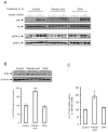
Figure 1
Figure 1
Palmitic acid but not DHA, inhibits insulin action and up-regulates TLR4. Human SH-Y5Y neuroblastoma cells were pretreated with palmitic acid, DHA or placebo during 4 h, then insulin-dependent Akt and ERK1/2 phosphorylation measured by Western blots using adequate antibodies, Control, Palmitic acid and DHA blots were performed in separate membranes, in each case the same membrane was blotted with different antobodies (panel A); TLR4 expression was measured by Western blot using anti-TLR4 antibodies and the expression was normalized to β tubilin, and band densities were quantified and expressed as ratio of TLR4/β-tubilin (panel B); TLR4 mRNA expression was determined by qRT-PCR normalized to GAPDH (panel C). Western blot and qRT-PCR data were presented as means ± SEM (n = 3), * and ** denoted significant differences vs control at p < 0.05 and p < 0.005, respectively.
Figure 2
Figure 2
Palmitic acid but not DHA, increased the expression of pro-inflammatory factors. (Panel A) human SH-SY5Y neuroblastoma cells stably tranfected with NF-κB luciferase reporter gene were treated with palmitic acid, DHA or placebo, and then relative luciferase activity measured. (Panel B) Human SH-SY5Y neuroblastoma cells were treated with palmitic acid, DHA or placebo and then IL6 and TNFα expression was determined using adequate primers, and normalized using GAPDH. Data were presented as means ± SEM (n = 3), * and ** denoted significant differences vs control at p < 0.05 and p < 0.005, respectively.
Figure 3
Figure 3
Resistin increased neuroinflammation through its binding to TLR4. (Panel A) Immunoprecipitation/ Immunoblot (IP/IB) analysis of the direct association of resistin with TLR4 in protein extracts from SH-SY5Y cells treated with resistin (100 ng/ml) for 16 h in the presence or absence of the cross-linker agent BS3, the shown blots are from the same membrane but blotted with different antibodies following immunoprecipitation with anti-TLR4 antibody. (Panel B,C) SH-SY5Y cells were treated with or without resistin and then Akt and p38 MAP kinase phosphorylation measured by Western blots using adequate antibodies and the presented blots was from the same membrane but blotted successively with different antibodies, the band densities were quantified and expressed as phosphorylated/total proteins. Data were presented as means ± SEM (n = 3), * and *** denoted significant differences vs control at p < 0.05 and p < 0.0005, respectively. (Panel D) human SH-SY5Y neuroblastoma cells stably transfected with NF-κB luciferase reporter gene were treated resistin and relative luciferase activity determined. (Panel F) Human SH-SY5Y neuroblastoma cells were treated with resistin in the presence or absence of Akt, Erk, JNK or p38 MAP kinase inhibitors, and then IL6 and TNFα expression was determined using specific primers for these two genes, and normalized using GAPDH. Data were presented as means ± SEM (n = 3). Columns marked by different letter differ (with or without asterix concerns TNFα or IL6, respectively) significantly (p < 0.05). For other panels, data were presented as means ± SEM (n = 3), * and *** denoted significant differences at p < 0.05 and p < 0.005, respectively. (Panel E) Human SH-SY5Y neuroblastoma cells were treated with resistin or placebo and then IL6 and TNFα expression was determined using adequate primers, and normalized using GAPDH. Data were presented as means ± SEM (n = 3), * and *** denoted significant differences at p < 0.05 and p < 0.001, respectively. (Panel F).
Figure 4
Figure 4
Palmitic acid promoted resistin action. Human SH-Y5Y cells were pretreated with palmitic acid (panel A) or DHA (panel B) and then stimulated by resistin. Phosphorylated Akt and p38 MAP kinase was determined by Western blot using adequate antibodies. (Panel C) cells were treated with DHA or placebo and p38 MAP kinase phosphorylation was determined by Western blot using adequate antibodies. The presented blots in (panel A), (panel B) and (panel C) are from the same membranes that were successively blotted with different antibodies. (Panel D) human SH-SY5Y neuroblastoma cells stably transfected with NF-κB luciferase reporter gene were treated resistin, resistin + palmitic acid or resistin + DHA, and relative luciferase activity determined. Data were presented as means ± SEM (n = 3), ** and *** denoted significant differences vs control at p < 0.005 and p < 0.001, respectively. (Panel E) Human SH-SY5Y neuroblastoma cells were treated with resistin, resistin + palmitic acid or resistin + DHA, and then IL6 and TNFα expression was determined using adequate primers, and normalized using GAPDH. Data were presented as means ± SEM (n = 3), * and ** denoted significant differences vs control at p < 0.05 and p < 0.005, respectively.
Figure 5
Figure 5
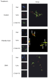
Palmitic acid induced TLR4 recruitment to membrane lipid rafts. Human SH-SY5Y cells were treated with palmitic acid, DHA or placebo during 4 h and fixed for immunohistochemistry analysis by confocal microscopy using antibodies directed towards CTXB (marker of membrane lipid rafts) or TLR4. The arrow in the merge indicated the co-localization of TLR4 and CTXB. Images were captured using a confocal microscope with high-magnification scan (X40). The figure is representative of two biological replicates and each image resulted from several slides that were analyzed by confocal microscopy.
Figure 6
Figure 6
Palmitic acid required TLR4 to partially impair insulin signaling and induced inflammation. (Panel A) Human SH-SY5Y cells were transiently transfected with siTLR4 or scramble (control siRNA) and then TLR4 expression was measured by RT-qPCR. Data were presented as means ± SEM (n = 3). *** denoted p < 0.001 when comparing control siRNA to TLR4siRNA, statistical analysis was performed using Student t-test. (Panel B) Human SH-SY5Y cells were transiently transfected with siTLR4 or scramble (control siRNA), cells were cultured in the presence or absence of palmitic acid and then TNFα expression measured by RT-qPCR. Data were presented as means ± SEM (n = 3). * and ** denoted p < 0.05 and p < 0.01, respectively, when comparing control vs palmitate for both cells exposed to control or palmitate condition, statistical analysis was performed using Student t-test. (Panel C) Human SH-SY5Y cells transfected with control siRNA (scramble) or siTLR4 were preatreated with or without palmitic acid during 4 h then acutely stimulated by insulin. The upper part of panel C shows representative Western blots using anti-pAkt, anti-p-ERK1/2 or anti-αTubilin antibodies and each antibody was used in different membranes. The lower part of panel C shows band density quantification. Phosphorylated Akt and Erk1/2 were normalized to αTubilin. Data were presented as means ± SEM (n = 3). # and ## denoted significant differences at p < 0.05 and p < 0.01, respectively when comparing insulin-dependent phosphorylation of Akt and Erk1/2 between W/O pretreatment and palmitate treatment groups, statistical analysis was performed using Kruskal Wallis test and post-hoc Pairwise comparison in R. ** and *** denoted significant differences at p < 0.05, p < 0.1 and p < 0.001 when comparing Control vs Insulin, statistical analysis was performed using Student t-test.
Figure 7
Figure 7
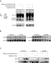
Palmitic acid increased resistin-dependent TLR4/MYD88/TIRAP association. (Panel A) Immunoprecipitation/Immunoblot (IP/IB) showing the association of TLR4 with MyD88 and TIRAP was analyzed in human SH-SY5Y neuroblastoma cells following treatment with palmitic acid or DHA and in response to resistin by Western blot using adequate antibodies. All blots are from the same membrane. Each experiment has been performed in triplicates as technical replicates. (Panel B) Human SH-SY5Y cells were transfected with scramble (Control siRNA), siTLR4 or siMyD88 and then TLR4 and MyD88 expression were measured by Western blot normalized to actin, for each antibody different blots were used. Each experiment has been performed in triplicates as technical replicates. (Panel C) Palmitic acid, resistin or placebo either treated these transfected cells and then Western blots were performed using anti-pAkt or Akt. These blots were performed in independent membranes. Each experiment has been performed in triplicates as technical replicates.

Similar articles

Cited by

References

    1. Schenk S, Saberi M, Olefsky JM. Insulin sensitivity: modulation by nutrients and inflammation. J. Clin. Invest. 2008;118:2992–3002. doi: 10.1172/JCI34260. - DOI - PMC - PubMed
    1. Ilievski V, Cho Y, Katwala P, Rodriguez H, Tulowiecka M, Kurian D, Leoni L, Christman JW, Unterman TG, Watanabe K. TLR4 expression by liver resident cells mediates the development of glucose intolerance and insulin resistance in experimental periodontitis. PLoS ONE. 2015;10(8):e0136502. doi: 10.1371/journal.pone.0136502. - DOI - PMC - PubMed
    1. Zheng C, Yang Q, Xu C, Shou P, Cao J, Jiang M, Chen Q, Cao G, Han Y, Li F, Cao W, Zhang L, Shi Y, Wang Y. CD11b regulates obesity-induced insulin resistance via limiting activation and proliferation of adipose tissue macrophages. Proc. Natl. Acad. Sci. USA. 2015;112:E7239–E7248. doi: 10.1073/pnas.1500396113. - DOI - PMC - PubMed
    1. Kain V, Kapadia B, Viswakarma N, Seshadri S, Prajapati B, Jena PK, Meda CLT, Subramanian M, Suraj SK, Kumar ST, Babu PP, Thimmapaya B, Reddy JK, Parsa KVL, Misra P. Co-activator binding protein PIMT mediates TNFa induced insulin resistance in skeletal muscle via the transcriptional down-regulation of MEF2A and Glut4. Sci. Rep. 2015;5:15197. doi: 10.1038/srep15197. - DOI - PMC - PubMed
    1. Dey D, Bhattacharya A, Roy S, Bhattacharya S. Fatty acid represses insulin receptor gene expression by impairing HMGA1 through protein kinase Cepsilon. Biochem. Biophys. Res. Commun. 2007;357:474–479. doi: 10.1016/j.bbrc.2007.03.183. - DOI - PubMed

Publication types