. 2021 Apr;40(14):2553-2566.
doi: 10.1038/s41388-020-01599-z. Epub 2021 Mar 8.

RGS12 is a novel tumor suppressor in osteosarcoma that inhibits YAP-TEAD1-Ezrin signaling

Affiliations

RGS12 is a novel tumor suppressor in osteosarcoma that inhibits YAP-TEAD1-Ezrin signaling

Yang Li et al. Oncogene. 2021 Apr.

Abstract

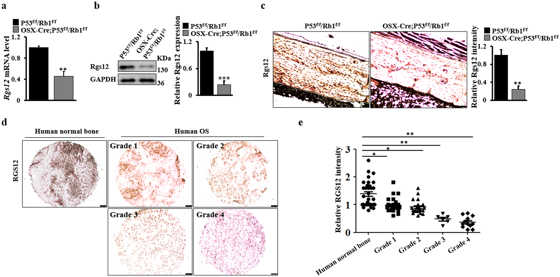
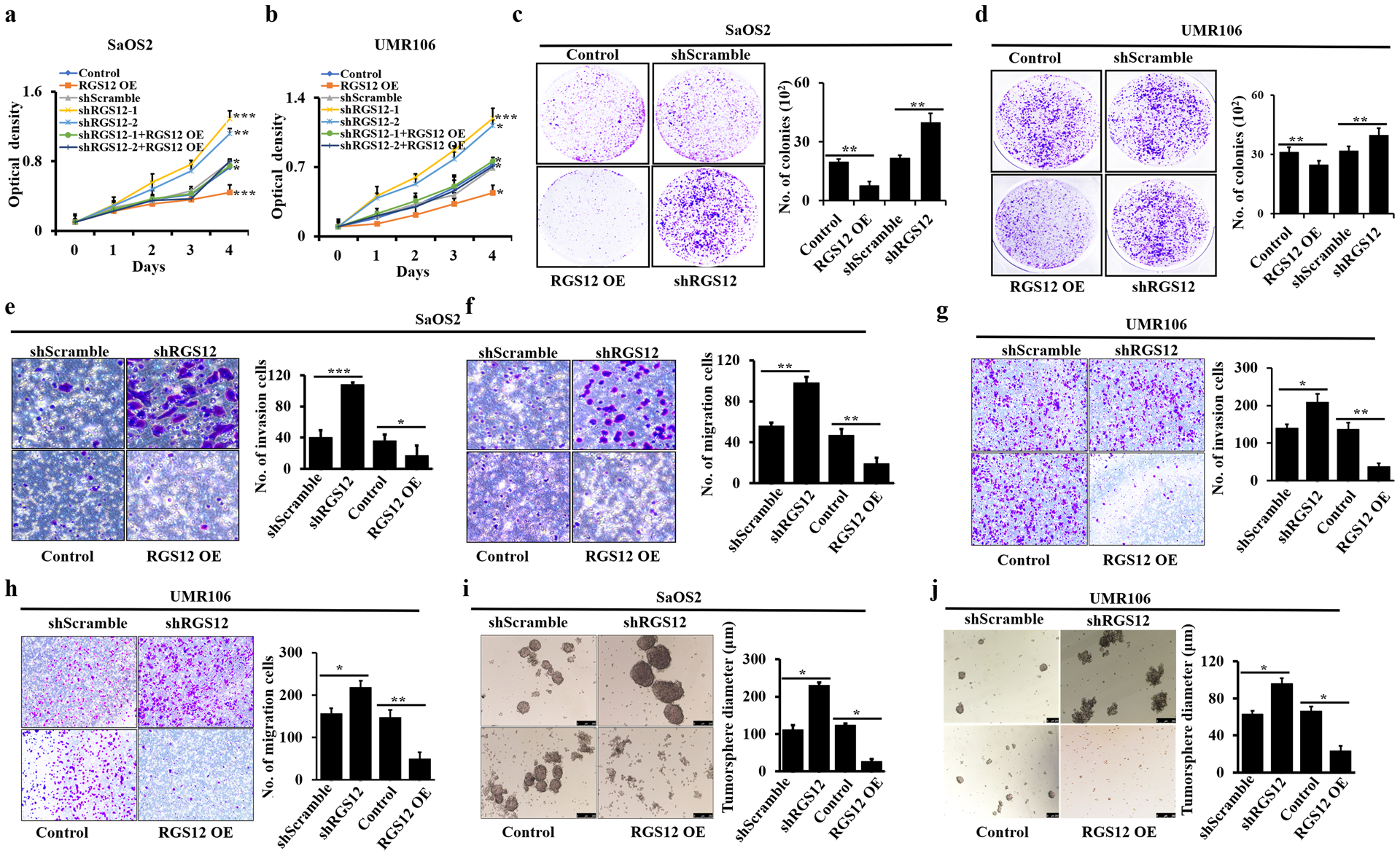
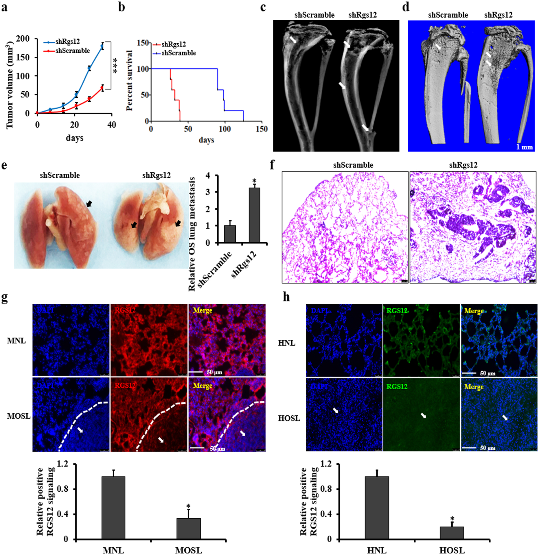
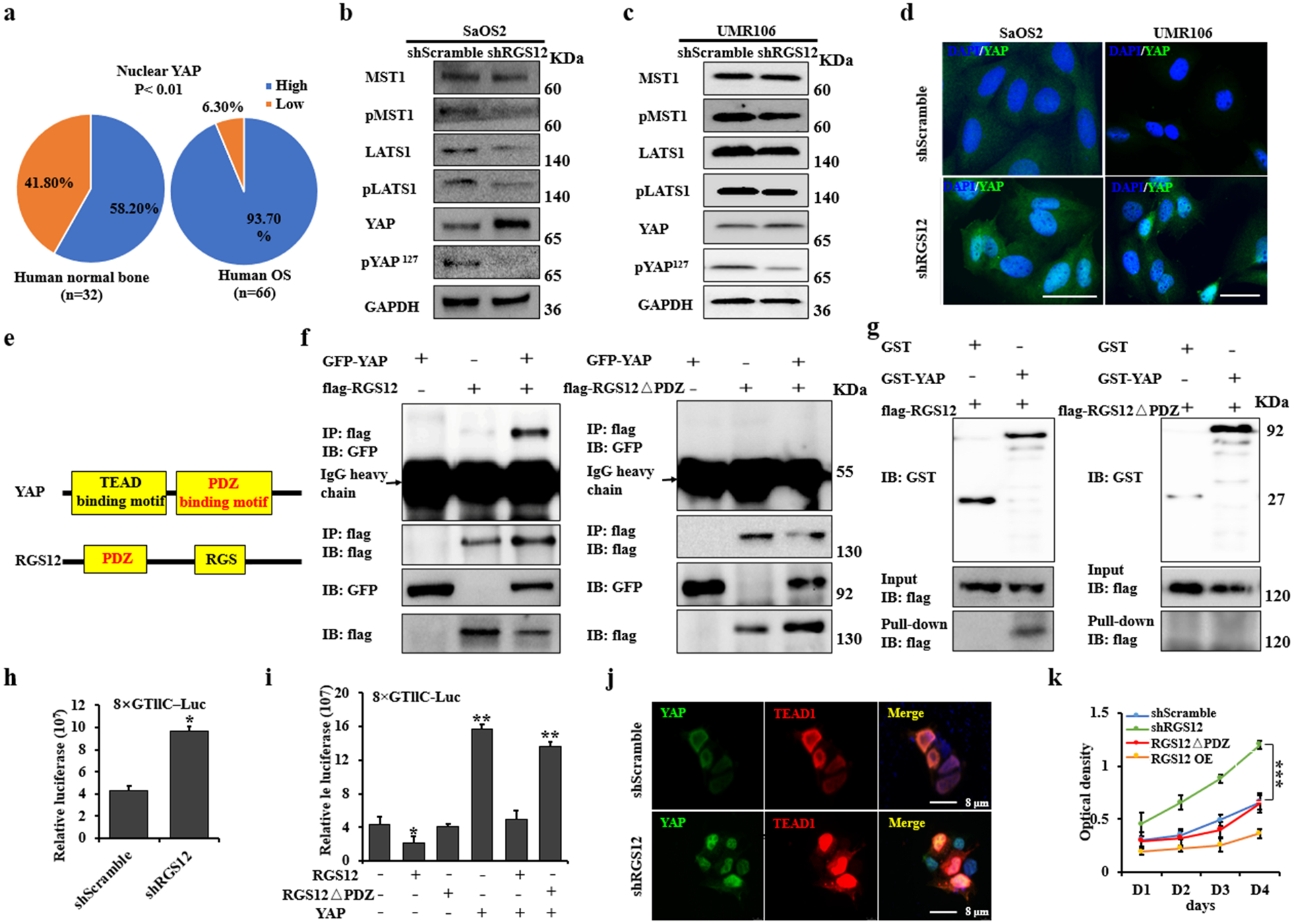
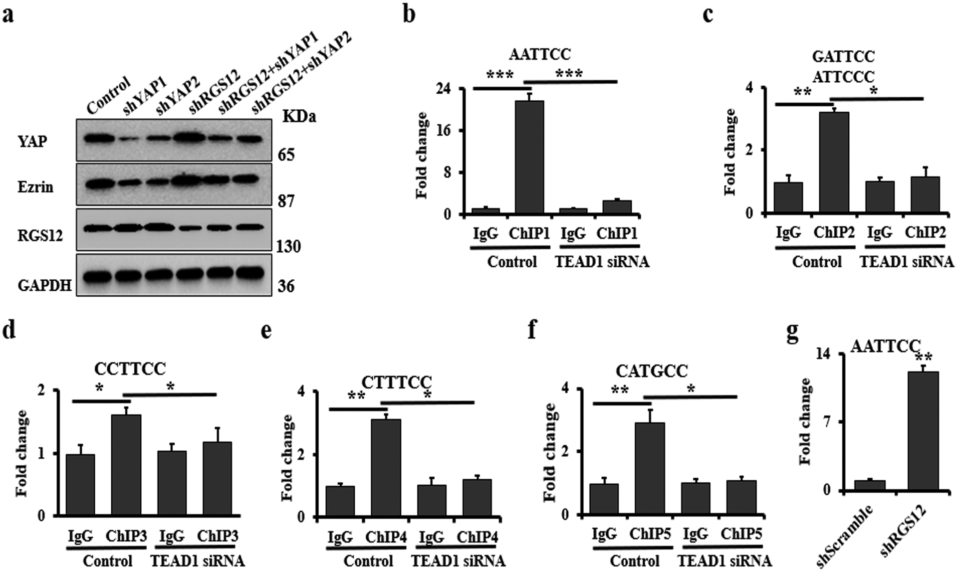
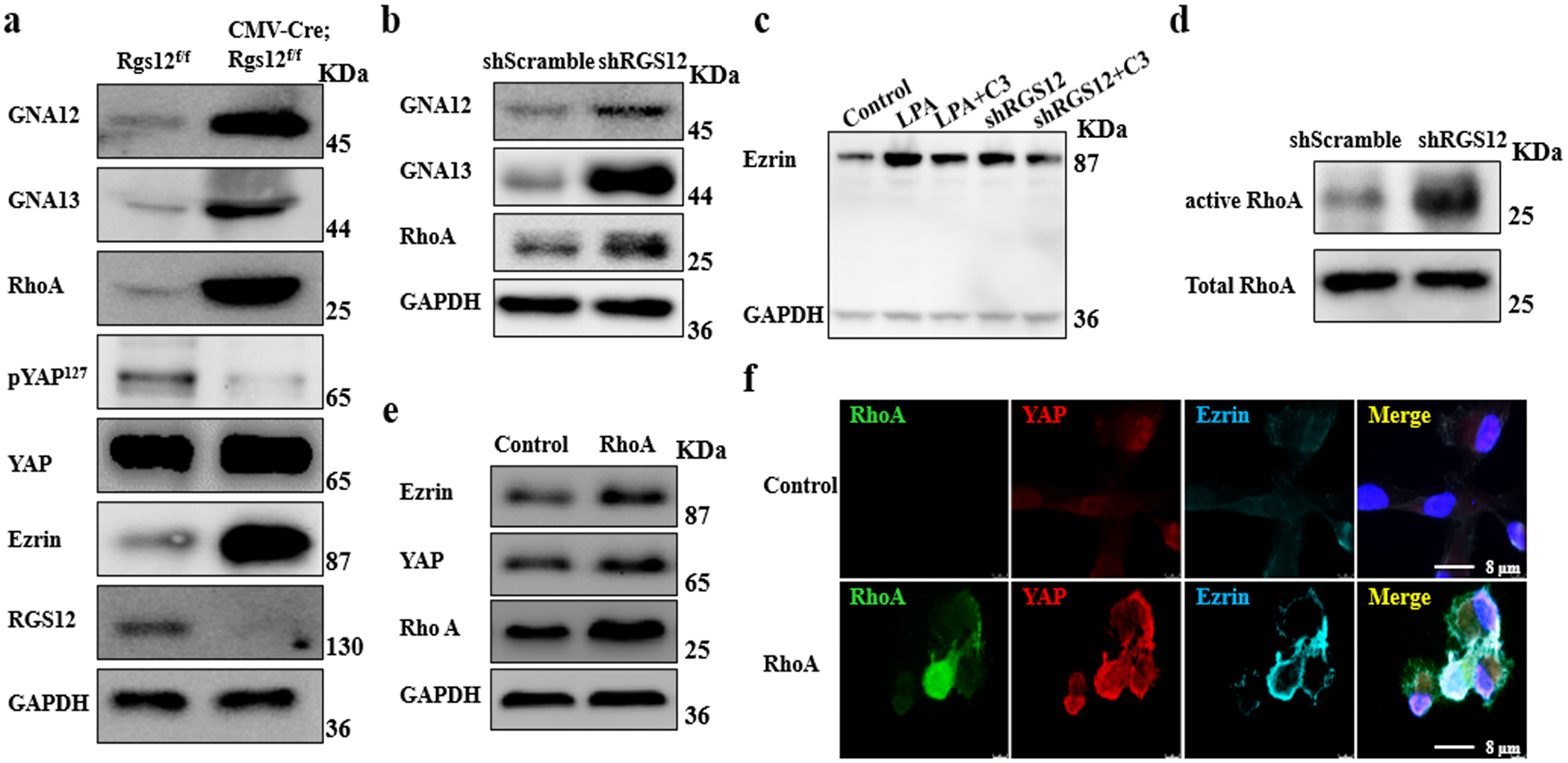
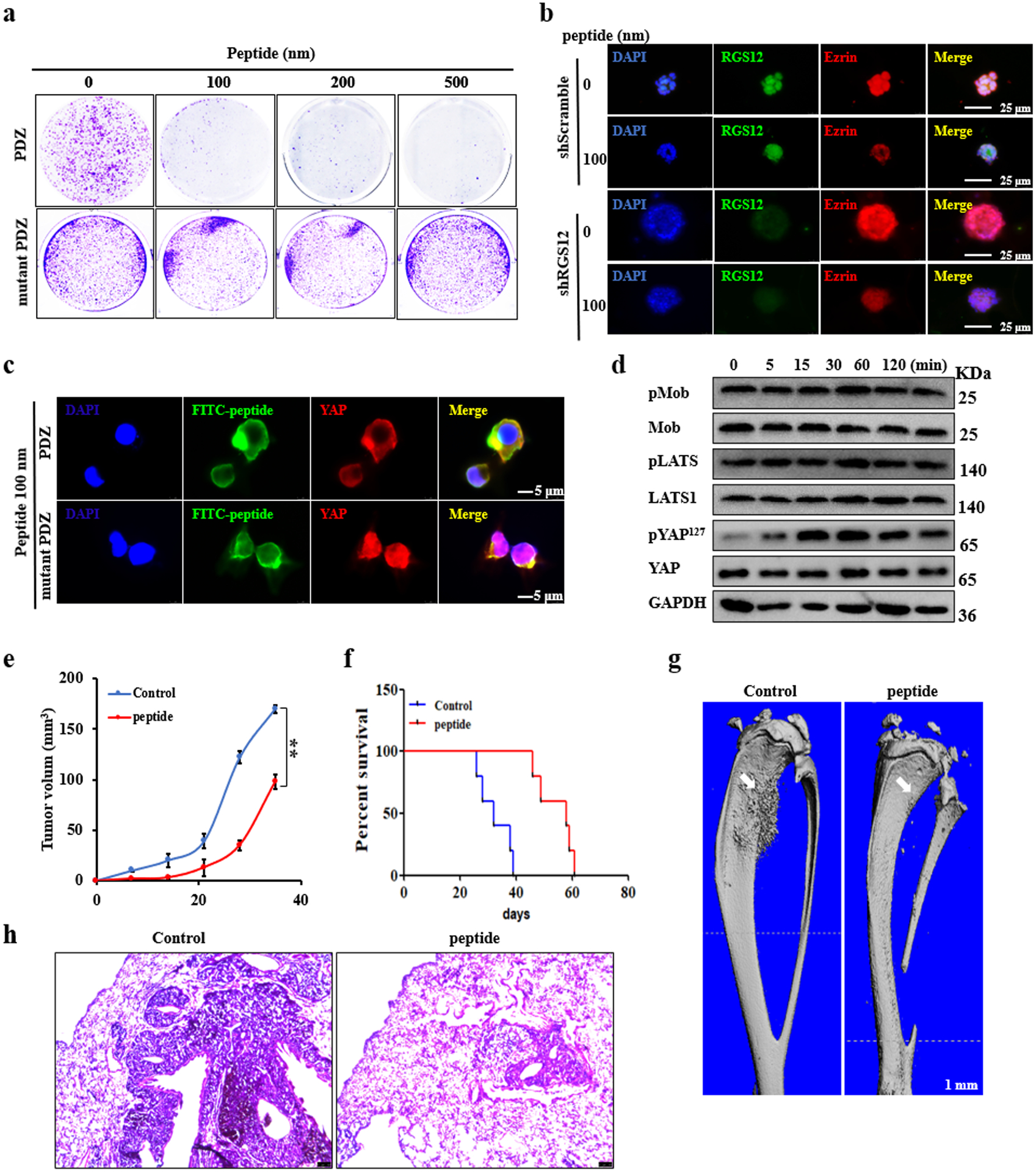
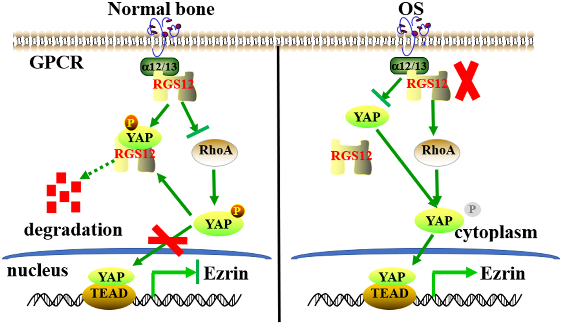
Osteosarcoma (OS) is the most common primary malignancy of the bone that predominantly affects children and adolescents. Hippo pathway is a crucial regulator of organ size and tumorigenesis. However, how Hippo pathway regulates the occurrence of osteosarcoma is largely unknown. Here, we reported the regulator of G protein signaling protein 12 (RGS12) is a novel Hippo pathway regulator and tumor suppressor of osteosarcoma. Depletion of Rgs12 promotes osteosarcoma progression and lung metastasis in an orthotopic xenograft mouse model. Our data showed that the knockdown of RGS12 upregulates Ezrin expression through promoting the GNA12/13-RhoA-YAP pathway. Moreover, RGS12 negatively regulates the transcriptional activity of YAP/TEAD1 complex through its PDZ domain function to inhibit the expression and function of the osteosarcoma marker Ezrin. PDZ domain peptides of RGS12 can inhibit the development of intratibial tumor and lung metastases. Collectively, this study identifies that the RGS12 is a novel tumor suppressor in osteosarcoma through inhibiting YAP-TEAD1-Ezrin signaling pathway and provides a proof of principle that targeting RGS12 may be a therapeutic strategy for osteosarcoma.


Conflict of interest statement

Conflict of interest

The authors declare no competing interests.

Figures

Fig. 1.
Fig. 1.. RGS12 is downregulated in both human and mouse osteosarcoma tissues.
a Real-time PCR quantification of Rgs12 mRNA levels in the bones of OSX-Cre;P53f/f/Rb1f/f and P53f/f/Rb1f/f mice. b A representative image of western blot. Whole bone lysates from OSX-Cre;P53f/f/Rb1f/f and P53f/f/Rb1f/f mice were immunoblotted with antibodies against Rgs12 and GAPDH, respectively. N=3. The Rgs12 expression is quantified by ImageJ software in the corresponding column at right. c A representative image of immunohistochemical staining of Rgs12 on mouse osteosarcoma and normal bones. Scale bar, 75 μm. N=5. The Rgs12 expression is quantified by ImageJ software in the corresponding column at right. d A representative image of immunohistochemical staining of RGS12 on different grades of human osteosarcoma (tumor grades 1 to 4) and normal bones. Scale bar, 100 μm. OS, osteosarcoma. e, Quantification of RGS12 expression by ImageJ software based on IHC staining in human osteosarcoma (tumor grades 1 to 4; Grade 1, N=27; Grade 2, N=21; Grade 3, N=6; Grade 4, N=12) and normal bones (N=32). Error bars were the means ± standard error of the mean (SEM) of triplicates from a representative experiment. *P < 0.05, **P < 0.01 and ***P < 0.001.
Fig. 2.
Fig. 2.. RGS12 inhibits osteosarcoma cell migration, invasion and tumorsphere formation in vitro.
a-b The cell proliferation rates of indicated cells were detected by WST-1 assay. vs. shScramble or normal control cells. c, d The analyses of colony formation abilities in the indicated cells. Cell numbers are quantified in the corresponding column at right. e, g Cell invasion. Cell numbers are quantified in the corresponding column at right. f, h Cell migration. Cell numbers are quantified in the corresponding column at right. i-j Tumorspheres. Error bars were the means ± standard error of the mean (SEM) of triplicates from a representative experiment. *P < 0.05, **P < 0.01 and ***P < 0.001.
Fig. 3.
Fig. 3.. Knockdown of RGS12 in SaOS2 promotes intratibial primary tumor growth and lung metastasis in SCID mice.
a Primary intratibial tumor growth over time in individual mice injected with shScramble or shRGS12 cells. b Kaplan-Meier survival analysis indicating the overall survival of mice injected with shScramble or shRGS12 cells. c, d Representative X-ray and micro-CT images of tumor-bearing legs after intratibial injection with shScramble or with shRGS12 cells at Day 28. The white arrow indicates the tumor in the bone. e Representative images of osteosarcoma lung metastasis of mice injected with shScramble or shRGS12 cells. Osteosarcoma lung metastasis is quantified in the corresponding column at right. The black arrow indicates the tumor in the lung. f Representative images of HE-stained lung sections of mice injected with shScramble or shRGS12 cells at Day 28. Scale bar, 100 μm. g Representative images of immunofluorescent staining of RGS12 in osteosarcoma lung metastasis sections of mice injected with shScramble or shRGS12 cells. The white arrow indicates the tumor in the lung. RGS12+ signaling is quantified in the corresponding column at lower panel. MNL, mouse normal lung tissues; MOSL, mouse osteosarcoma lung metastasis tissues. h Representative images of immunofluorescence staining for RGS12 in human osteosarcoma lung metastasis sections and controls. RGS12+ signaling is quantified in the corresponding column at lower panel. HNL, human normal lung tissues; HOSL, human osteosarcoma lung metastasis tissues. Error bars are the means ± standard error of the mean (SEM) of triplicates from a representative experiment. *P < 0.05 and ***P < 0.001.
Fig. 4.
Fig. 4.. RGS12 inhibits transcriptional YAP/TEAD1 activity through its PDZ domain function.
a Statistical analysis of nuclear YAP expression in human osteosarcoma and normal bone specimens. b, c Whole protein lysates of shScramble and shRGS12 cells were immunoblotted with the indicated antibodies. d Representative immunofluorescence-stained images of shScramble and shRGS12 cells for YAP expression, DAPI staining for nuclear. e Structures of YAP and RGS12. f Co-IP experiments of GFP-YAP, flag-RGS12 or flag-RGS12ΔPDZ in 293T cells. g 293T cells were transfected with flag-RGS12 or flag-RGS12ΔPDZ, respectively. Cells were lysed after 48 hr, and cell lysates were incubated with GST or GST-YAP protein on glutathione beads. The precipitated complexes were analyzed by western blot. h Scramble and shRGS12 cells were respectively seeded in 12-well plates. Luciferase reporter and pRL-TK vector (internal control) were co-transfected. Luciferase activities were measured after transfection of 48 hr. i Luciferase activity was measured in SaOS2 cells following co-transfection with flag-YAP, flag-RGS12 or flag-RGS12ΔPDZ, respectively. j Immunofluorescent staining of YAP and TEAD1 in shScramble and shRGS12 cells. k The proliferation of the indicated cells was detected by WST-1 assay. Error bars were the means ± standard error of the mean (SEM) of triplicates from a representative experiment. *P < 0.05, **P < 0.01, ***P < 0.001.
Fig. 5.
Fig. 5.. Knockdown of RGS12 induces YAP-dependent Ezrin expression.
a Immunoblots of the whole protein lysates, which were isolated from the cells after co-transfection with shRGS12 or/both shYAP1/2 lentivirus for 48 hrs. b-f ChIP. Co-occupation of YAP/TEAD1 using YAP antibody in the Ezrin promoter. c ChIP. Co-occupation of YAP/TEAD1 using YAP antibody in the Ezrin promoter after silence of RGS12. Error bars were the means ± standard error of the mean (SEM). *P < 0.05, **P < 0.01, ***P < 0.001.
Fig. 6.
Fig. 6.. RGS12 negatively regulates Ezrin expression via GNA12/13-RhoA-YAP pathway
a Bone tissues from CMV-Cre;Rgs12f/f and control mice were analyzed by immunoblotting with the indicated antibodies. b Western blot for analyzing GNA12, GNA13 and RhoA expression in the lysates from shScramble and shRGS12 cells. c SaOS2 cells were starved for 12 hr and then treated with 2 mg/mL C3 (RhoA inhibitor) or 1 mM LPA (GPCR activator) for 48 hr. The whole protein lysates of the treated cells were analyzed by western blot. d RhoA activity. e Western blot analysis of the whole protein lysates isolated from the SaOS2 cells that were transfected with empty vector (Control) and pcDNA3.1-RhoA plasmid, respectively. f Immunofluorescent staining of RhoA, Ezrin and YAP in SaOS2 cells that were transfected with empty vector (Control) and pcDNA3.1-RhoA plasmid, respectively.
Fig. 7.
Fig. 7.. RGS12 PDZ peptides inhibit osteosarcoma formation and lung metastasis.
a Soft agar. Colony formation ability analysis after PDZ or PDZ mutated peptide treatment for 3 weeks in SaOS2 cells. b Immunofluorescent staining of RGS12 and Ezrin in shScramble and shRGS12 cells after co-culture of PDZ peptides and matrigel for 7 days. c, d PDZ peptides inhibited YAP nuclear translocation by increasing the phosphorylation level of YAP in SaOS2 cells. e Representative X-ray images of tumor-bearing legs after intratibial injection with shRGS12 cells mixed with 0 or 100 nm PDZ peptides. f Kaplan-Meier survival analysis indicating overall survival of mice injected with shRGS12 cells mixed with 0 or 100 nM of PDZ peptides. g Representative micro-CT images of tumor-bearing legs after intratibial injection with shRGS12 cells mixed with 0 or 100 nM of PDZ peptides at Day 35. The white arrow indicates the tumor in the bone. h Representative images of HE-stained lung sections of mice injected with shRGS12 cells mixed with 0 or 100 nM of PDZ peptides at Day 35. Scale bar, 100 μm. Error bars were the means ± standard error of the mean (SEM) of triplicates from a representative experiment. **P < 0.01.
Fig. 8.
Fig. 8.. A proposed model to illustrate the regulatory mechanism of RGS12 on Ezrin expression in osteosarcoma.
In normal bone, RGS12 promotes the growth arrest of osteosarcoma through inhibiting YAP nuclear translocation. In osteosarcoma bone, RGS12 has a lower expression and knockdown of RGS12 enhances RhoA activity and the transcriptional activity of YAP/TEAD1 to induce Ezrin expression.

References

    1. Kansara M, Teng MW, Smyth MJ, Thomas DM. Translational biology of osteosarcoma. Nat Rev Cancer 2014; 14: 722–735. - PubMed
    1. Tang QL, Lu JC, Zou CY, Shao Y, Chen Y, Narala S et al. CDH4 is a novel determinant of osteosarcoma tumorigenesis and metastasis. Oncogene 2018; 37: 3617–3630. - PubMed
    1. Wang DY, Wu YN, Huang JQ, Wang W, Xu M, Jia JP et al. Hippo/YAP signaling pathway is involved in osteosarcoma chemoresistance. Chin J Cancer 2016; 35: 1–8. - PMC - PubMed
    1. Harvey KF, Zhang XM, Thomas DM. The Hippo pathway and human cancer. Nat Rev Cancer 2013; 13: 246–257. - PubMed
    1. Yu FX, Guan KL. The Hippo pathway: regulators and regulations. Genes & development 2013; 27: 355–371. - PMC - PubMed

Publication types

MeSH terms