Review
. 2021 Feb;113(1):54-65.
doi: 10.32074/1591-951X-234.

Histopathology of Non-IBD Colitis. A practical approach from the Italian Group for the study of the gastrointestinal tract (GIPAD)

Affiliations
Review

Histopathology of Non-IBD Colitis. A practical approach from the Italian Group for the study of the gastrointestinal tract (GIPAD)

Vincenzo Villanacci et al. Pathologica. 2021 Feb.

Abstract

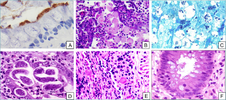
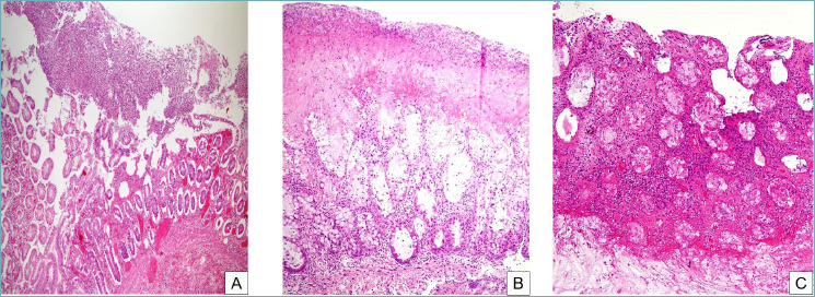
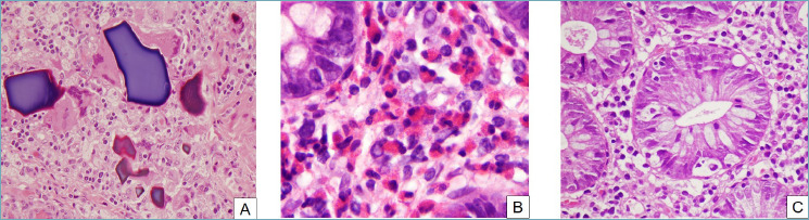
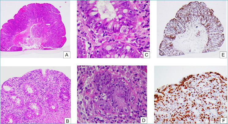
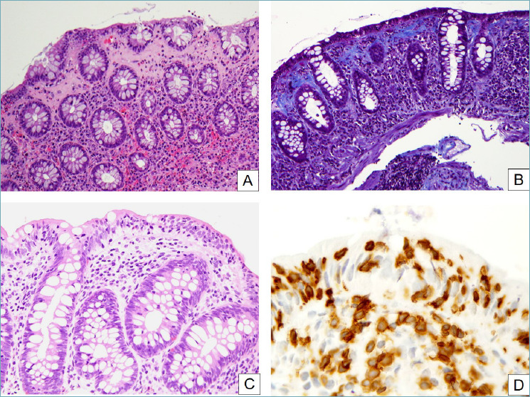
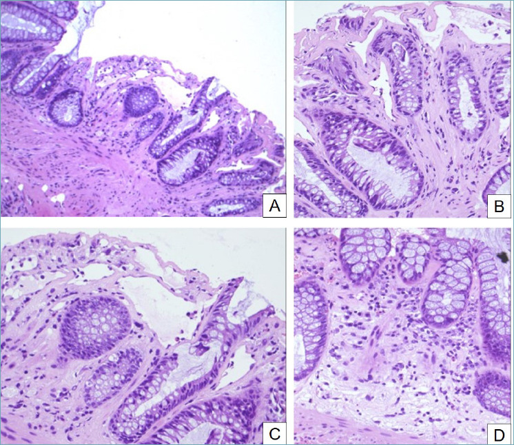
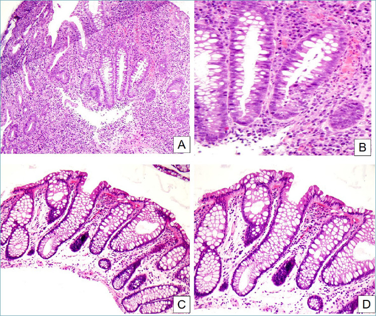
Non-IBD colitides (NIBDC) are intestinal diseases clinically and endoscopically overlapping with Inflammatory Bowel Diseases (IBD), sometimes with a similar histological picture. NIBDC include entities such as infectious colitis, ischemic colitis, pseudomembranous colitis, eosinophilic colitis, autoimmune enterocolitis, segmental colitis associated with diverticulosis, drug-induced colitis, radiation-induced colitis, diversion colitis, and microscopic colitis, this last including two entities: collagenous and lymphocytic colitis. The knowledge of the most useful histological features and the main clinical data for each entity is mandatory in daily clinical practice, for correct pathological diagnosis and clinical management.

Keywords: colitis; drug-induced colitis; infectious colitis; ischemic colitis; microscopic colitis.


Conflict of interest statement

Conflict of interest

The Authors declare no conflict of interest.

Figures

Figure 1.
Figure 1.
Infectious colitis. (A) Intestinal spirochetosis. The fuzzy luminal border is highlighted by immunohistochemistry for Treponema pallidum (peroxidase-diaminobenzidine, 40x). (B) Entamoeba histolytica trophozoites containing engulfed erythrocytes can be seen among neutrophils and inflammatory debris (H&E, 20x). (C) Grocott’s stain makes Histoplasma capsulatum stand out as intensely-stained small oval structures within the cytoplasm of macrophages (Grocott’s methenamine silver stain, 20x). (D) Strongyloides stercoralis can be found lurking in the crypts (H&E, 20x). (E) CMV-infected cells stand out as markedly enlarged cells with typical “owl’s eye” intranuclear inclusions (H&E, 20x). (F) Cryptosporidium parvum forms small round bodies on the luminal border of enterocytes (H&E, 20x). !
Figure 2.
Figure 2.
Pseudomembranous colitis. (A) At low magnification, a mushroom-shaped necroinflammatory exudate overlies an acutely injured mucosa (H&E, 10x). (B) The pseudomembrane is composed of inflammatory cells, necrotic debris, mucus and fibrin. Cryptitis and crypt abscesses are evident in the mucosa (H&E, 20x). (C) The severity of the inflammation can vary widely, from a mild picture to severe and deep acute inflammation which can obscure other morphological features (H&E, 20x).
Figure 3.
Figure 3.
Drug-induced colitis. Various histological patterns. (A) Drug crystals and remnants can sometimes be seen in a colonic biopsy. In some cases, such as this one, they are surrounded by a foreign-body reaction (H&E, 20x). (B) Eosinophilic colitis can be caused by drugs. In this picture, numerous eosinophils infiltrate the lamina propria and crypt epithelium, forming microabscesses and sometimes degranulating (H&E, 40x). (C) Prominent apoptotic bodies can be seen in some cases of drug-induced colitis, especially with some laxatives and antineoplastic agents (H&E, 40x).
Figure 4.
Figure 4.
Autoimmune enteropathy. (A) At low power the mucosa appears reactive, with regenerative crypts and an hypercellular lamina propria (H&E, 2x). (B) Crypts are regenerative, with mucin depletion and surface epithelial damage (H&E, 10x). (C) On closer inspection, there are prominent apoptotic bodies in the surface and crypt epithelium (H&E, 40x). (D) Sometimes, satellite lymphocytes can be seen around the apoptotic bodies (H&E, 40x). (E) CD3 immunohistochemistry reveals a greatly increased T lymphocyte population (H&E, 2x). (F) The T lymphocytes can be seen in the lamina propria as well as in crypt and surface epithelium (H&E, 20x).
Figure 5.
Figure 5.
Microscopic colitis. (A) In collagenous colitis, a thick and irregular collagen band underlies the surface epithelium and often entraps inflammatory cells. Surface epithelial detachment, shown here, is also typical. The underlying lamina propria is inflamed and shows an increased amount of eosinophils (H&E, 20x). (B) A Masson trichrome clearly highlights the irregularly thickened subepithelial collagen band (Masson trichrome, 20x). (C) In lymphocytic colitis, numerous T lymphocytes infiltrate the gland epithelium (H&E, 20x). (D) CD3 stain clearly highlights the pathologically increased intraepithelial lymphocytes (peroxidase-diaminobenzidine, 40x).
Figure 6.
Figure 6.
Radiation-induced colitis. (A) Crypt distortion, dilation and atrophy are evident (H&E, 10x). (B) Fibrosis of lamina propria, hyperplasia of crypts and dilated vascular channels (H&E, 20x). (C) Crypt atrophy, apoptotic bodies, goblet cell depletion and ectasia of small vessels are typical of radiation-induced colitis (H&E, 20x). (D) The lamina propria shows an inflammatory infiltrate that tends to decrease in long-standing cases (H&E, 20x).
Figure 7.
Figure 7.
Segmental colitis associated with diverticular disease. (A) In this case, crypt distortion, cryptitis and basal plasmacytosis dominate the picture (H&E, 10x). (B) Detail of the previous image. Active inflammation and cryptitis can be appreciated, as well as the increased number of plasma cells and eosinophils in the inflammatory infiltrate (H&E, 40x). (C, D) In this other case, inflammation is much less florid, while crypt distortion and stromal edema are predominant (H&E, 20x and 40x respectively).

Similar articles

Cited by

References

    1. Schofield JB, Haboubi N. Histopathological mimics of inflammatory bowel disease. Inflamm Bowel Dis 2020. June 18;26(7):994-1009. https://doi.org/10.1093/ibd/izz232 10.1093/ibd/izz232 - DOI - PubMed
    1. Villanacci V, Reggiani-Bonetti L, Caprioli F, et al. . Histopathology of inflammatory bowel disease - Position statement of the Pathologists of the Italian Group for the Study of Inflammatory Bowel Disease (IG-IBD) and Italian Group of Gastrointestinal Pathologists (GIPAD-SIAPEC). Dig Liv Dis 2020;52:262-7. https://doi.org/10.1016/j.dld.2019.11.005 10.1016/j.dld.2019.11.005 - DOI - PubMed
    1. Villanacci V, Reggiani-Bonetti L, Salviato T, et al. . Histopathology of IBD Colitis. A practical approach from the pathologists of the Italian Group for the study of the gastrointestinal tract (GIPAD). Pathologica 2021;113:39-53. https://doi.org/10.32074/1591-951X-235 10.32074/1591-951X-235 - DOI - PMC - PubMed
    1. Leder K, Torresi J, Brownstein JS, et al. . Travel-associated illness trends and clusters, 2000-2010. Emerg Infect Dis 2015;19:1049-73. https://doi.org/10.3201/eid1907.121573 10.3201/eid1907.121573 - DOI - PMC - PubMed
    1. Lamps LW. Update on infectious enterocolitis and the diseases that they mimic. Histopathology 2015;66:3-14. https://doi.org/10.1111/his.12582 10.1111/his.12582 - DOI - PubMed

LinkOut - more resources