Review
. 2021 Mar 12;135(5):703-724.
doi: 10.1042/CS20200331.

Mechanisms and clinical manifestations of cardiovascular toxicities associated with immune checkpoint inhibitors

Affiliations
Review

Mechanisms and clinical manifestations of cardiovascular toxicities associated with immune checkpoint inhibitors

Alan H Baik et al. Clin Sci (Lond). .

Abstract

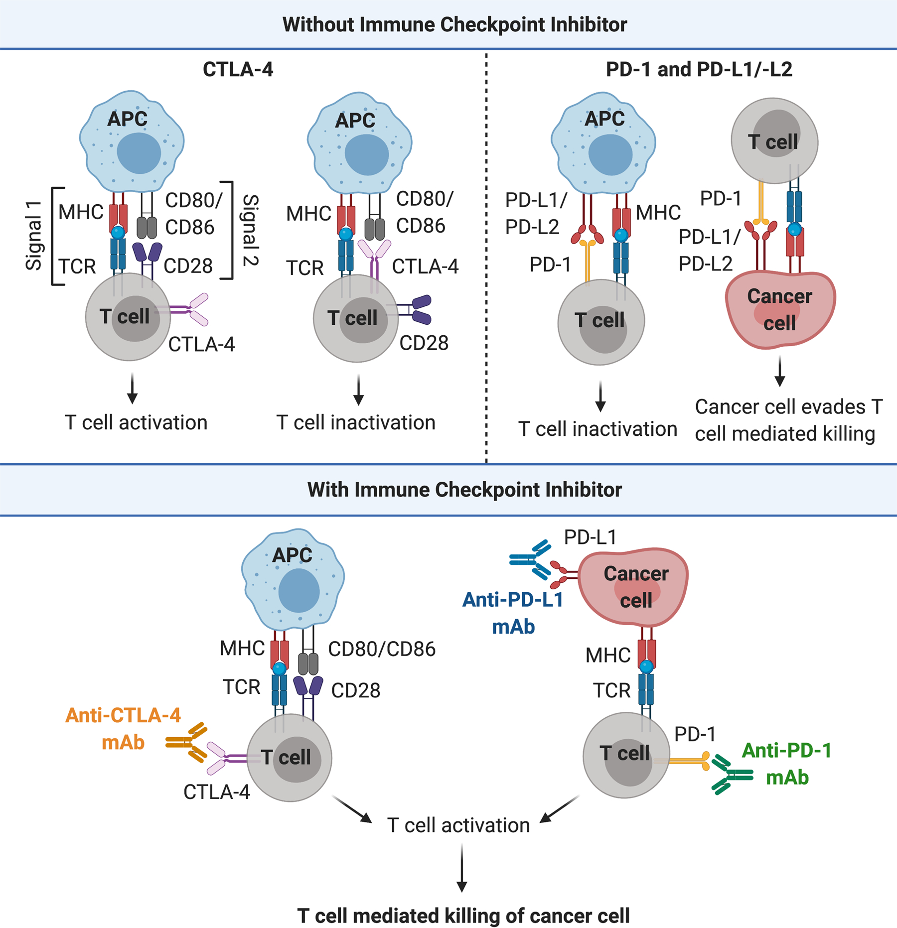
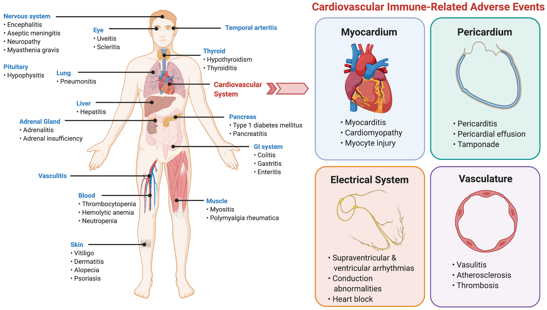
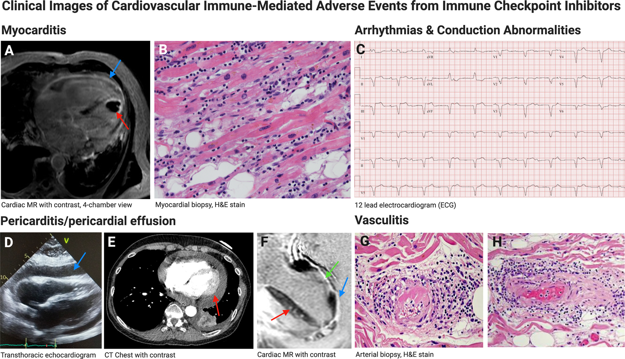
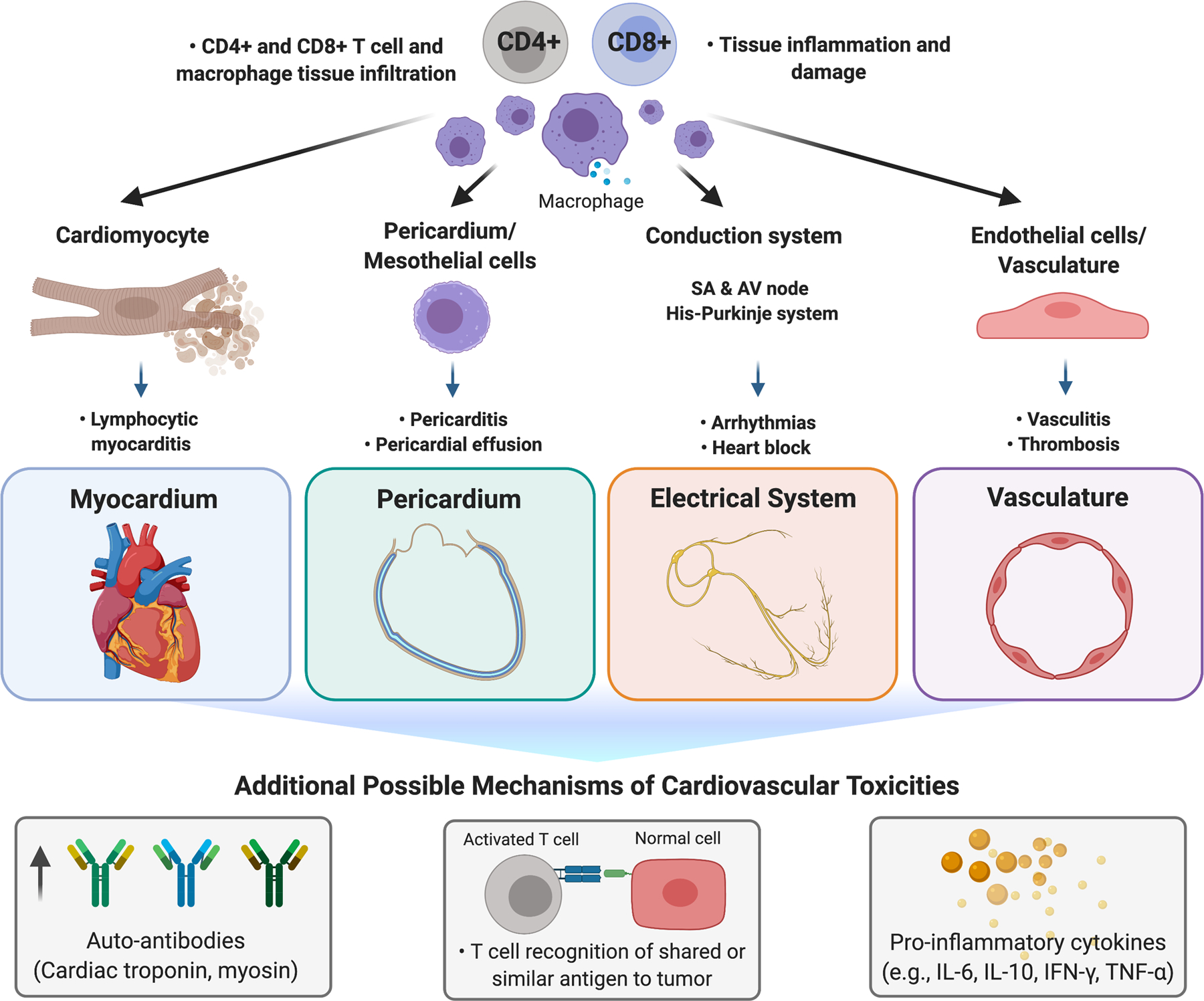
Immunotherapies have greatly expanded the armamentarium of cancer-directed therapies in the past decade, allowing the immune system to recognize and fight cancer. Immune checkpoint inhibitors (ICIs), in particular, have revolutionized cancer treatment and have demonstrated survival benefit in numerous types of cancer. These monoclonal antibodies increase anti-cancer immunity by blocking down-regulators of adaptive immunity, including cytotoxic T lymphocyte-associated protein 4 (CTLA-4), programmed cell death protein 1 (PD-1), and its ligand (PD-L1), resulting in anti-tumor activity. As ICIs increase immune system activation, they can cause a wide range of inflammatory side effects, termed immune-released adverse events. Though these toxicities can affect nearly any organ, the most fatal toxicity is myocarditis. Here, we discuss the diverse spectrum of cardiovascular toxicities associated with ICI use. In addition, we provide insight and future directions on mechanisms and treatments for immune-related adverse events (irAEs) involving the myocardium, pericardium, vasculature, and conduction system.

Keywords: autoimmunity; cardiovascular disease; heart failure; immunomodulation.


Figures

Figure 1:
Figure 1:
Immune checkpoints and immune checkpoint inhibitors. CTLA-4 and PD-1/PD-L1/2 signaling dampen T cell activation to maintain a balance between immune activation and self-tolerance. Immune checkpoint inhibitors block CTLA-4 and PD-1/PD-L1 signaling, leading to T cell activation and T cell-mediated killing of cancer cells. Abbreviations: APC, antigen presenting cell; CD, cluster of differentiation; CTLA-4, cytotoxic T lymphocyte-associated protein 4; MHC, major histocompatibility complex; PD-1, programmed cell death protein 1; TCR, T cell receptor
Figure 2:
Figure 2:
Types of immune checkpoint inhibitor-associated immune-related adverse events. Immune checkpoint inhibitors can cause a wide range of inflammatory side effects called immune-related adverse events (irAEs). These toxicities can affect nearly every organ system. Cardiovascular system irAE can involve the myocardium, pericardium, vasculature, and conduction system.
Figure 3:
Figure 3:. Clinical images of ICI-associated cardiovascular toxicities.
Myocarditis. A) Cardiac MR with contrast 4-chamber view, showing thinning of left ventricle with subendocardial and subepicardial/pericardial late gadolinium enhancement (blue arrow) and thrombus in the left ventricle (red arrow). B) Photomicrograph of endomyocardial biopsy (H&E staining) demonstrating prominent lymphocytic interstitial inflammation, interstitial edema, and severe injured myocytes with pyknotic nuclei and hyper-eosinophilic cytoplasm (400x). Arrhythmias and Conduction Abnormalities. C) Electrocardiogram showing left bundle branch block and third-degree heart block. Pericardial Toxicities. D) Transthoracic echocardiogram, subcostal view, showing large, circumferential pericardial effusion (blue arrow). E) CT chest showing large pericardial effusion with pericardial enhancement suggestive of pericarditis (red arrow). F) Cardiac MR with contrast, 4-chamber view showing diffuse pericardial enhancement (blue arrow), transmural late gadolinium enhancement in mid-lateral wall (green arrow) and patchy mid-septal wall enhancement (red arrow). Vasculitis. G, H) Photomicrographs of artery biopsy (H&E staining) demonstrating necrotizing vasculitis and perivascular lymphocytic infiltration. Myocarditis H&E image courtesy of Dr. Javid Moslehi, Vanderbilt University. Vasculitis H&E images courtesy of Dr. Robert Padera, Harvard Medical School.
Figure 4:
Figure 4:
Mechanisms of immune checkpoint inhibitor associated cardiovascular toxicity. ICI-associated myocarditis is characterized by extensive lymphocytic infiltration (CD4+ T cells, CD8+ T cells, CD68+ macrophages) into the myocardium, myocyte injury, and myonecrosis. Hypothetical mechanisms include increased auto-antibodies that target self-antigens (e.g., cardiac troponin, myosin), T cell recognition of a shared or similar antigen between the tumor and normal cells, and elevation of pro-inflammatory cytokines.

Similar articles

Cited by

References

    1. Esensten JH, Helou YA, Chopra G, Weiss A, and Bluestone JA, “CD28 Costimulation: From Mechanism to Therapy,” Immunity, vol. 44, no. 5, pp. 973–988, 17 2016, doi: 10.1016/j.immuni.2016.04.020. - DOI - PMC - PubMed
    1. Schildberg FA, Klein SR, Freeman GJ, and Sharpe AH, “Coinhibitory Pathways in the B7-CD28 Ligand-Receptor Family,” Immunity, vol. 44, no. 5, pp. 955–972, 17 2016, doi: 10.1016/j.immuni.2016.05.002. - DOI - PMC - PubMed
    1. Romo-Tena J, Gómez-Martín D, and Alcocer-Varela J, “CTLA-4 and autoimmunity: new insights into the dual regulator of tolerance,” Autoimmun Rev, vol. 12, no. 12, pp. 1171–1176, October. 2013, doi: 10.1016/j.autrev.2013.07.002. - DOI - PubMed
    1. Francisco LM, Sage PT, and Sharpe AH, “The PD-1 pathway in tolerance and autoimmunity,” Immunol. Rev, vol. 236, pp. 219–242, July. 2010, doi: 10.1111/j.1600-065X.2010.00923.x. - DOI - PMC - PubMed
    1. Ribas A and Wolchok JD, “Cancer immunotherapy using checkpoint blockade,” Science, vol. 359, no. 6382, pp. 1350–1355, 23 2018, doi: 10.1126/science.aar4060. - DOI - PMC - PubMed

Publication types

MeSH terms

Substances