. 2021 Mar 3;13(6):8127-8145.
doi: 10.18632/aging.202618. Epub 2021 Mar 3.

Long noncoding RNA NKILA transferred by astrocyte-derived extracellular vesicles protects against neuronal injury by upregulating NLRX1 through binding to mir-195 in traumatic brain injury

Affiliations

Long noncoding RNA NKILA transferred by astrocyte-derived extracellular vesicles protects against neuronal injury by upregulating NLRX1 through binding to mir-195 in traumatic brain injury

Bin He et al. Aging (Albany NY). .

Abstract

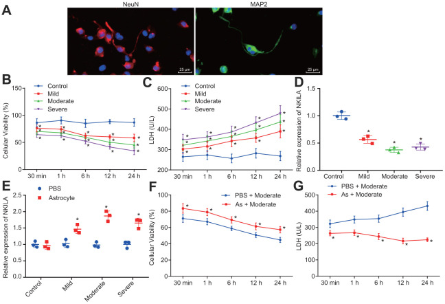
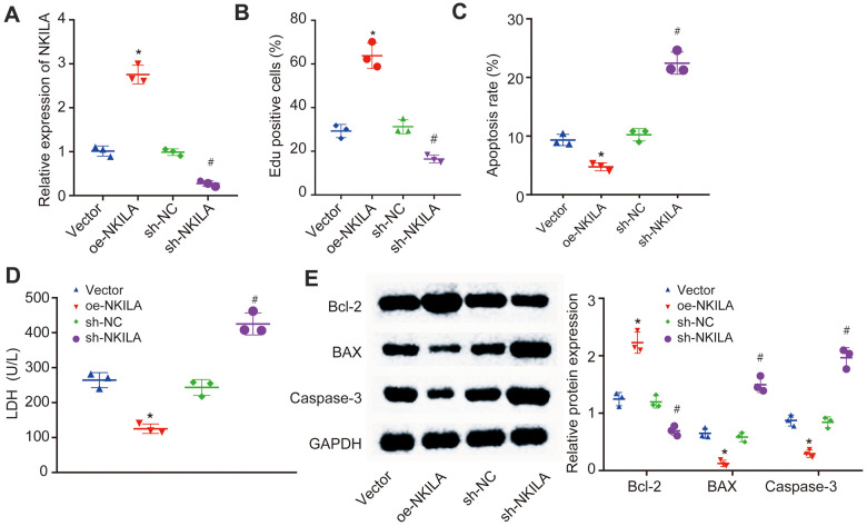
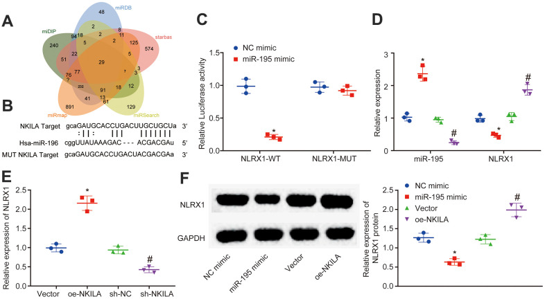
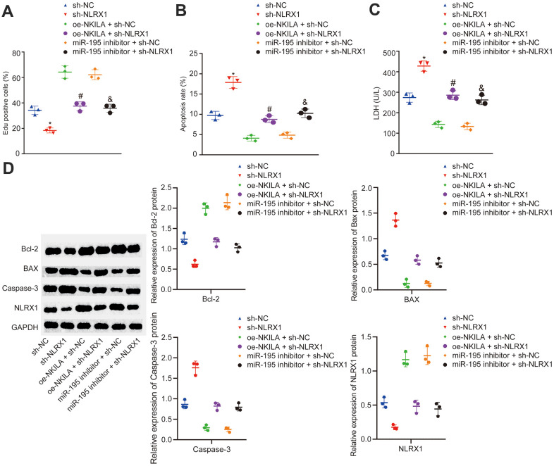
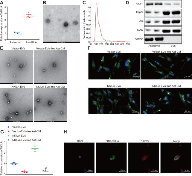
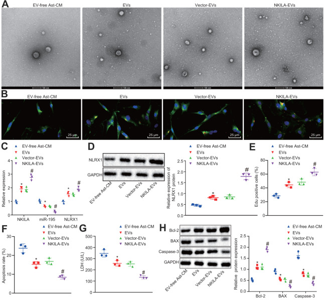
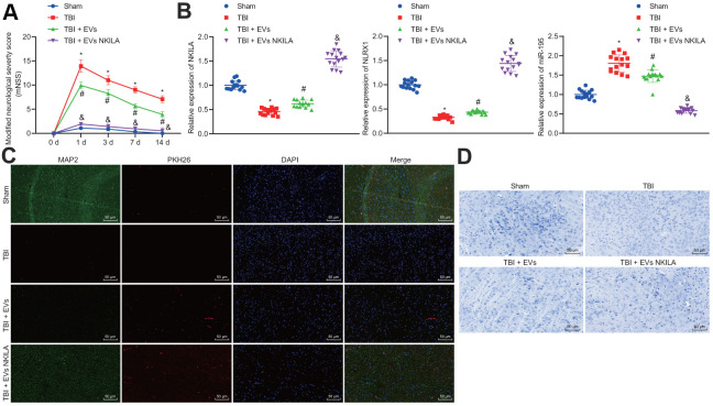
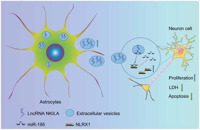
The study aims to investigate the effects of long noncoding RNA (lncRNA) transmitted nuclear factor-κB interacting lncRNA (NKILA)-containing astrocyte-derived small extracellular vesicles (EVs) on traumatic brain injury (TBI). TBI was modeled in vitro by exposing human neurons to mechanical injury and in vivo by controlled cortical impact in a mouse model. The gain- and loss-function approaches were conducted in injured neurons to explore the role of NKILA, microRNA-195 (miR-195) and nucleotide-binding leucine-rich repeat containing family member X1 (NLRX1) in neuronal injury. EVs extracted from NKILA-overexpressing astrocytes were used to treat injured neurons. It was revealed that NKILA was downregulated in injured neurons. Astrocyte co-culture participated in the upregulation of NKILA in injured neurons. Additionally, NKILA could competitively bind to miR-195 that directly targeted NLRX1. Next, the upregulation of NLRX1 or NKILA relived neuronal injury by promoting neuronal proliferation but inhibiting apoptosis. Astrocyte-derived EVs transferred NKILA into neurons, which led to the downregulation of miR-195, upregulation of NLRX1, increased cell proliferation, and decreased cell apoptosis. The in vivo experiments validated that NKILA-containing EVs promoted brain recovery following TBI. Collectively, astrocyte-derived EVs carrying NKILA was found to alleviate neuronal injury in TBI by competitively binding to miR-195 and upregulating NLRX1.

Keywords: NLRX1; extracellular vesicles; long noncoding RNA NKILA; microRNA-195; traumatic brain injury.


Conflict of interest statement

CONFLICTS OF INTEREST: The authors declare that they have no conflicts of interest.

Figures

Figure 1
Figure 1
Astrocytes upregulates NKILA to promote recovery of injured neurons. (A) immunochemical staining of MAP2 and NeuN expression in neurons (× 400). (B) proliferation of neurons with mild, moderate or severe injury detected by CCK-8 assay. (C) LDH content in the culture medium of neurons with mild, moderate or severe injury. (D) NKILA expression in neurons with mild, moderate or severe injury detected by RT-qPCR. (E) NKILA expression in injured neurons after co-culture of astrocytes detected by RT-qPCR. (F) neuronal proliferation in injured neurons after co-culture of astrocytes detected by CCK-8 assay. (G) LDH content in injured neuron after co-culture of astrocytes. All data were measurement data and expressed as mean ± standard deviation. Independent sample t test was used for comparison between two groups (E). The one-way ANOVA was used for comparison among multiple groups, followed by Tukey’s post-hoc test (D) and two-way ANOVA for comparisons between time-based measurements within each group (B, C, F, G). All experiments were done at least three independent times. * p < 0.05 compared with control neurons or moderately injured neurons cultured with PBS.
Figure 2
Figure 2
NKILA promotes proliferation and inhibits apoptosis of injured neurons. The injured neurons were introduced with oe-NKILA, sh-NKILA, vector or sh-NC. (A) NKILA expression in injured neurons after treatment measured by RT-qPCR. (B) cell proliferation of injured neurons after treatment measured by EdU assay. (C) quantitative analysis for cell apoptosis of injured neurons after treatment measured by flow cytometry. (D) LDH content in injured neurons after treatment. (E) protein expression of apoptosis-related factors (Bcl-2, Bax and Caspase-3) in injured neurons after treatment measured by Western blot analysis. * p < 0.05 compared with neurons treated with vector; # p < 0.05 compared with neurons treated with sh-NC. All data were measurement data and expressed as mean ± standard deviation. One-way ANOVA was used for comparison among multiple groups, followed by Tukey’s post-hoc test. The cell experiment was repeated three times.
Figure 3
Figure 3
NKILA competitively binds to miR-195. (A) the subcellular location of NKILA in neurons predicted by the lncATLAS website. (B) the subcellular localization of NKILA in neurons detected by FISH assay (× 400). (C) the potential binding sites between NKILA and miR-195 predicted by RNA22. (D) the binding relationship between NKILA and miR-195 measured by dual-luciferase reporter gene assay. * p < 0.05 compared with the treatment of vector. (E) the enrichment of miR-195 caused by NKILA detected by RNA pull-down. * p < 0.05 compared with the treatment of Bio-probe NC. (F) the binding of NKILA and miR-195 with AGO2 determined by RIP assay. * p < 0.05 compared with IgG. All data were measurement data and expressed as mean ± standard deviation. Unpaired t test was used for pairwise comparison in panel D and F, and one-way ANOVA followed by Tukey’s post-hoc test was used for comparison in panel E. The cell experiment was repeated three times.
Figure 4
Figure 4
NKILA acts as ceRNA of miR-195 to upregulate NLRX1. (A) Venn analysis of target genes of miR-195 obtained from miDIP, miRDB and starbase. (B) putative binding sites between NLRX1 and miR-195 predicted in RNA22. (C) the binding relationship between NLRX1 and miR-195 verified by dual-luciferase reporter gene assay. * p < 0.05 compared with the treatment of NC mimic. (D) NLRX1 and miR-195 expression in neurons after alteration of miR-195 detected by RT-qPCR. * p < 0.05 compared with the treatment of NC mimic. # p < 0.05 compared with the treatment of NC inhibitor. (E) NLRX1 mRNA expression in neurons after alteration of NKILA detected by RT-qPCR. * p < 0.05 compared with the treatment of vector. # p < 0.05 compared with the treatment of sh-NC. (F) NLRX1 protein expression in neurons after overexpression of NKILA or miR-195 measured by Western blot analysis. * p < 0.05 compared with the treatment of NC mimic. # p < 0.05 compared with the treatment of vector. All data were measurement data and expressed as mean ± standard deviation. Unpaired t test was used for comparison between two groups. The one-way ANOVA was used for comparison among multiple groups, followed by Tukey’s post-hoc test. The cell experiment was repeated three times.
Figure 5
Figure 5
NKILA promotes proliferation and inhibits apoptosis of injured neurons by increasing NLRX1 expression via miR-195. The injured neurons were introduced with oe-NKILA + sh-NC, oe-NKILA + sh-NLRX1, miR-195 inhibitor + sh-NC, miR-195 inhibitor + sh-NLRX1, sh-NLRX1 or sh-NC. (A) cell proliferation of injured neurons after treatment measured by EdU assay. (B) quantitative analysis for cell apoptosis of injured neurons after treatment measured by flow cytometry. (C) LDH content in injured neurons after treatment. (D) protein expression of NLRX1 and apoptosis-related factors (Bcl-2, Bax and Caspase-3) in injured neurons after treatment measured by Western blot analysis. * p < 0.05 compared with the treatment of sh-NC. # p < 0.05 compared with the treatment of oe-NKILA + sh-NC. & p < 0.05 compared with the treatment of miR-195 inhibitor + sh-NC. All data were measurement data and expressed as mean ± standard deviation. Unpaired t test was used for comparison between two groups. The one-way ANOVA was used for comparison among multiple groups, followed by Tukey’s post-hoc test. The cell experiment was repeated three times.
Figure 6
Figure 6
Astrocytes transfer NKILA into neurons through EVs. (A) NKILA expression in astrocytes detected by RT-qPCR. (B) representative electron micrograph of EVs isolated from astrocytes (scale bar: 100 nm). The red arrow points to the EVs. (C) size and particle distribution plots of isolated EVs from culture medium by NTA. (D) expression of GLT-1, Hsp70, CD63 and Alix as well as the negative marker GRP94 evaluated by Western blot analysis. (E) observation of EVs released by astrocytes under an electron microscope (scale bar: 100 nm). (F) microscopic views of uptake of EVs by neurons (× 400). (G) the expression of NKILA in the supernatant of the astrocyte-derived EVs and EV-free Ast-CM by RT-qPCR; * p < 0.05 compared with the Vector-EVs. # p < 0.05 compared with the treatment of NKILA-EVs. (H) uptake of EVs by neurons observed by the confocal laser microscopy (scale bar: 10 μm). All data were measurement data and expressed as mean ± standard deviation. Unpaired t test was used for comparison between two groups. One-way ANOVA, followed by Tukey’s post-hoc test was used for multi-group comparison. The cell experiment was repeated three times.
Figure 7
Figure 7
NKILA delivered by astrocyte-derived EVs promotes neuron recovery by increasing NLRX1 expression via miR-195 in vitro. The injured neurons were treated with NKILA-EVs and vector-EVs. (A) observation of EVs released by astrocyte under an electron microscope (scale bar: 100 nm). (B) microscopic views of uptake of EVs by neurons (× 400). (C) expression of NKILA, miR-195 and NLRX1 in injured neurons measured by RT-qPCR. (D) protein expression of NLRX1 in injured neurons measured by Western blot analysis. (E) cell proliferation of injured neurons after treatment measured by EdU assay. (F) quantitative analysis for cell apoptosis of injured neurons after treatment measured by flow cytometry. (G) LDH content in injured neurons after treatment. (H) expression of Bcl-2, Bax and Caspase-3 in injured neurons after treatment measured by Western blot analysis. * p < 0.05 compared with neurons co-cultured with EV-free Ast-CM. # p < 0.05 compared with neurons co-cultured with Vector-EVs. All data were measurement data and expressed as mean ± standard deviation. Unpaired t test was used for comparison between two groups. One-way ANOVA, followed by Tukey’s post-hoc test, was used for multi-group comparisons. The cell experiment was repeated three times.
Figure 8
Figure 8
Astrocyte-derived EVs loaded with NKILA promote brain recovery in TBI mice in vivo. Mice were randomly classified into sham-operated mice, TBI mice, TBI mice treated with EVs or NKILA-enriched EVs (15 mice/group). (A) the mNSS determined before TBI and on the 1, 3, 7 and 14 days after TBI. (B) the levels of NKILA and NLRX1 in mouse left cerebral cortex tissue determined with RT-qPCR. (C) the expression of neuron marker MAP2 in PKH26-labeled EVs assayed with immunofluorescence assay (× 200). (D) The loss of neuron cells assessed using Nissl staining (× 200). * p < 0.05 compared with sham-operated mice. # p < 0.05 TBI mice. & p < 0.05 TBI mice treated with EVs. N = 15. All data were measurement data and expressed as mean ± standard deviation. One-way ANOVA was used for comparison among multiple groups, followed by Tukey’s post-hoc test. Comparisons between time-based measurements on neuronal function were determined with repeated measures ANOVA.
Figure 9
Figure 9
The molecular mechanism of astrocyte-secreted EVs enriched with NKILA in TBI. Astrocytes secrete EVs to stimulate NKILA expression in neurons, which upregulates NLRX1 by competitively binding to miR-195 and prevents neuronal injury following TBI.

References

    1. Andelic N. The epidemiology of traumatic brain injury. Lancet Neurol. 2013; 12:28–29. 10.1016/S1474-4422(12)70294-6 - DOI - PubMed
    1. Loane DJ, Faden AI. Neuroprotection for traumatic brain injury: translational challenges and emerging therapeutic strategies. Trends Pharmacol Sci. 2010; 31:596–604. 10.1016/j.tips.2010.09.005 - DOI - PMC - PubMed
    1. Cooper DJ, Rosenfeld JV, Murray L, Arabi YM, Davies AR, D’Urso P, Kossmann T, Ponsford J, Seppelt I, Reilly P, Wolfe R, and DECRA Trial Investigators, and Australian and New Zealand Intensive Care Society Clinical Trials Group. Decompressive craniectomy in diffuse traumatic brain injury. N Engl J Med. 2011; 364:1493–502. 10.1056/NEJMoa1102077 - DOI - PubMed
    1. Sofroniew MV, Vinters HV. Astrocytes: biology and pathology. Acta Neuropathol. 2010; 119:7–35. 10.1007/s00401-009-0619-8 - DOI - PMC - PubMed
    1. Linnerbauer M, Wheeler MA, Quintana FJ. Astrocyte crosstalk in CNS inflammation. Neuron. 2020; 108:608–22. 10.1016/j.neuron.2020.08.012 - DOI - PMC - PubMed

Publication types