

Identification of PSMD7 as a prognostic factor correlated with immune infiltration in head and neck squamous cell carcinoma
- PMID: 33687056
- PMCID: PMC7990087
- DOI: 10.1042/BSR20203829
Identification of PSMD7 as a prognostic factor correlated with immune infiltration in head and neck squamous cell carcinoma
Abstract
Background: Recurrent locally advanced or metastatic head and neck squamous cell carcinoma (HNSCC) is associated with dismal prognosis because of its highly invasive behavior and resistance to conventional intensive chemotherapy. The identification of effective markers for early diagnosis and prognosis is important for reducing mortality and ensuring that therapy for HNSCC is effective. Proteasome 26S subunit, non-ATPase 7 (PSMD7) is an ATP-independent component of the 19S regulatory subunit. The prognostic value of PSMD7 and the association with immune infiltration in HNSCC remains unclear.
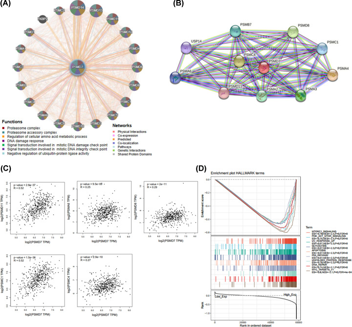
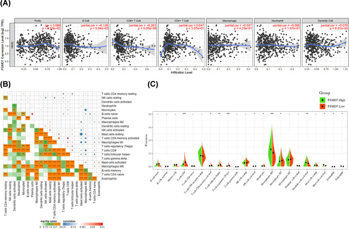
Methods: The Sangerbox, Oncomine, UALCAN and Human Protein Atlas (HPA) databases were used to examine PSMD7 expression profiles in HNSCC. The CVCDAP was used to analysis the association of PSMD7 with the prognosis of patients with HNSCC. The mechanism was investigated with gene set enrichment analysis (GSEA). The association between expression of PSMD7 and immune infiltration in HNSCC was investigated using the Tumor Immune Estimation Resource (TIMER), TISIDB database and CIBERSORT algorithm.
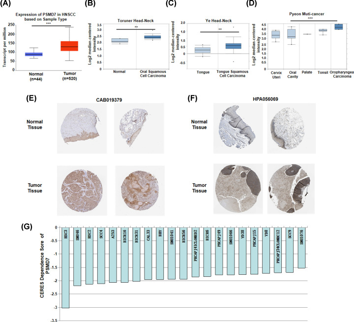
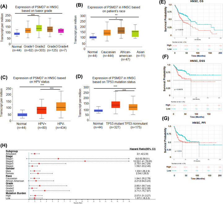
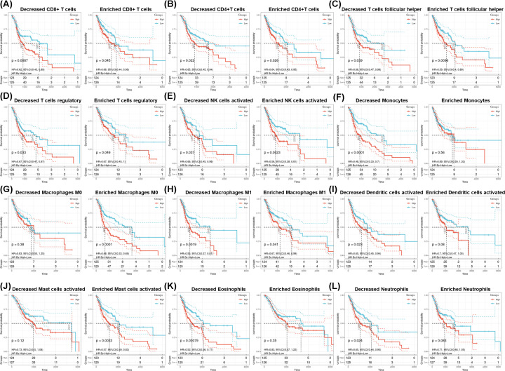
Results: PSMD7 expression was significantly up-regulated in HNSCC compared with relative normal tissues. In addition, up-regulated PSMD7 expression was associated with various clinicopathological parameters. High expression of PSMD7 suggested inferior survival of HNSCC patients. GSEA and CERES score indicated that PSMD7 was closely correlated with tumor-related signaling pathways and cell survival. Functional analyses revealed that PSMD7 was positively correlated with various infiltration levels. Moreover, PSMD7 influenced the prognosis of HNSCC patients partially via immune infiltration.
Conclusion: Our findings suggest that PSMD7 is associated poor prognosis in patients with HNSCC and plays an important role in tumor-related immune infiltration.
Keywords: HNSCC; OS; PSMD7; immune infiltration; prognostic factor.
© 2021 The Author(s).
Conflict of interest statement
The authors declare that there are no competing interests associated with the manuscript.
Figures
Similar articles
-
Identification of CCT3 as a prognostic factor and correlates with cell survival and invasion of head and neck squamous cell carcinoma.Biosci Rep. 2021 Oct 29;41(10):BSR20211137. doi: 10.1042/BSR20211137. Biosci Rep. 2021. PMID: 34505628 Free PMC article.
-
Overexpression of TPD52L2 in HNSCC: prognostic significance and correlation with immune infiltrates.BMC Oral Health. 2024 Oct 7;24(1):1191. doi: 10.1186/s12903-024-04977-1. BMC Oral Health. 2024. PMID: 39375696 Free PMC article.
-
CTHRC1 is a prognostic biomarker correlated with immune infiltration in head and neck squamous cell carcinoma.BMC Oral Health. 2024 Jun 27;24(1):742. doi: 10.1186/s12903-024-04525-x. BMC Oral Health. 2024. PMID: 38937712 Free PMC article.
-
Comprehensive review regarding the association of E2Fs with the prognosis and immune infiltrates in human head and neck squamous cell carcinoma.Asian J Surg. 2024 May;47(5):2106-2121. doi: 10.1016/j.asjsur.2024.01.130. Epub 2024 Feb 5. Asian J Surg. 2024. PMID: 38320907 Review.
-
Exploration of Feasible Immune Biomarkers for Immune Checkpoint Inhibitors in Head and Neck Squamous Cell Carcinoma Treatment in Real World Clinical Practice.Int J Mol Sci. 2020 Oct 15;21(20):7621. doi: 10.3390/ijms21207621. Int J Mol Sci. 2020. PMID: 33076306 Free PMC article. Review.
Cited by
-
19S Proteasome Subunits as Oncogenes and Prognostic Biomarkers in FLT3-Mutated Acute Myeloid Leukemia (AML).Int J Mol Sci. 2022 Nov 23;23(23):14586. doi: 10.3390/ijms232314586. Int J Mol Sci. 2022. PMID: 36498916 Free PMC article.
-
Deubiquitinase PSMD7 facilitates pancreatic cancer progression through activating Nocth1 pathway via modifying SOX2 degradation.Cell Biosci. 2024 Mar 17;14(1):35. doi: 10.1186/s13578-024-01213-9. Cell Biosci. 2024. PMID: 38494478 Free PMC article.
-
Proteomics Analysis of Aqueous Humor and Rejected Graft in Pig-to-Non-Human Primate Corneal Xenotransplantation.Front Immunol. 2022 Mar 24;13:859929. doi: 10.3389/fimmu.2022.859929. eCollection 2022. Front Immunol. 2022. PMID: 35401527 Free PMC article.
-
PSMD8 can serve as potential biomarker and therapeutic target of the PSMD family in ovarian cancer: based on bioinformatics analysis and in vitro validation.BMC Cancer. 2023 Jun 22;23(1):573. doi: 10.1186/s12885-023-11017-8. BMC Cancer. 2023. PMID: 37349676 Free PMC article.
-
Deubiquitinase PSMD7 promotes the proliferation, invasion, and cisplatin resistance of gastric cancer cells by stabilizing RAD23B.Int J Biol Sci. 2021 Jul 25;17(13):3331-3342. doi: 10.7150/ijbs.61128. eCollection 2021. Int J Biol Sci. 2021. PMID: 34512150 Free PMC article.
References
-
- de Mello R.A., Geros S., Alves M.P., Moreira F., Avezedo I. and Dinis J. (2014) Cetuximab plus platinum-based chemotherapy in head and neck squamous cell carcinoma: a retrospective study in a single comprehensive European cancer institution. PLoS ONE 9, e86697 10.1371/journal.pone.0086697 - DOI - PMC - PubMed
MeSH terms
Substances
LinkOut - more resources
Full Text Sources
Other Literature Sources
Medical