. 2021 Apr 7;12(7):1049-1060.
doi: 10.1021/acschemneuro.0c00719. Epub 2021 Mar 9.

Viral Involvement in Alzheimer's Disease

Affiliations

Viral Involvement in Alzheimer's Disease

Ahmad Sait et al. ACS Chem Neurosci. .

Abstract

Alzheimer's disease (AD) is characterized by the presence of β-amyloid plaques (Aβ) and neurofibrillary tangles (NFTs) in the brain. The prevalence of the disease is increasing and is expected to reach 141 million cases by 2050. Despite the risk factors associated with the disease, there is no known causative agent for AD. Clinical trials with many drugs have failed over the years, and no therapeutic has been approved for AD. There is increasing evidence that pathogens are found in the brains of AD patients and controls, such as human herpes simplex virus-1 (HSV-1). Given the lack of a human model, the route for pathogen entry into the brain remains open for scrutiny and may include entry via a disturbed blood-brain barrier or the olfactory nasal route. Many factors can contribute to the pathogenicity of HSV-1, such as the ability of HSV-1 to remain latent, tau protein phosphorylation, increased accumulation of Aβ invivo and in vitro, and repeated cycle of reactivation if immunocompromised. Intriguingly, valacyclovir, a widely used drug for the treatment of HSV-1 and HSV-2 infection, has shown patient improvement in cognition compared to controls in AD clinical studies. We discuss the potential role of HSV-1 in AD pathogenesis and argue for further studies to investigate this relationship.

Keywords: Alzheimer’s disease; apolipoprotein E; blood−brain barrier; herpes simplex virus; valacyclovir; β-amyloid.


Conflict of interest statement

The authors declare no competing financial interest.

Figures
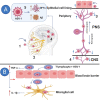
Figure 1
Figure 1
Possible routes for HSV-1 to enter the brain. Blue arrows indicate the virus movement. (A) (1) A suggested main route for HSV-1 to enter the brain is by infecting the epithelial cell line in the nasal cavity, causing a cold sore. (2) The virus can remain latent in the trigeminal nerve for an extended period of time. (3) Reactivation of the virus if immunocompromised and during chemotherapy. (4) The virus travels back to the primary site of infection and causes further cold sores and can reach the central nervous system (CNS) via sensory neurons in the peripheral nervous system (PNS). (5) HSV-1 induces the accumulation of β-amyloid plaques (Aβ) and neurofibrillary tangles (NFTs) inside the brain. (B) The virus can use T lymphocytes to cross the blood–brain barrier (BBB). The infection of the T lymphocyte with the virus stimulates the production of TNF-α and increases the production of interleukin-1β (IL-1β) in microglial cells, resulting in the breakdown of the BBB. Created with BioRender.com.
Figure 2
Figure 2
How HSV-1 might cause Alzheimer’s disease (AD). The main suggested routes to pathogenesis have been listed according to their ascending impact (from yellow, amber to red). The red sector shows biomarkers most prevalently associated with AD including Aβ accumulation, hyperphosphorylation of tau, and inflammation. These are related to HSV-1 studies where Aβ accumulation leads to AD through amyloid plaque formation. Hyperphosphorylation of TAU contributes to AD through paired helical filaments (PHFs) and neurofibrillary tangles (NFTs).
Figure 3
Figure 3
HSV 1 brain infection causes hyperphosphorylation of tau protein leading to Alzheimer’s disease. HSV-1 is able to increase CDK5, GSK3β, PKA, and kinase B activities while decreasing the activities of several phosphatases. These enzymes phosphorylate tau proteins (that normally stabilizes microtubules) at positions S214 and T212, among others. Microtubule structure is lost with phosphorylated tau creating cellular disorder. HSV-1 activates cdc2 kinase blocking cellular mitosis. Then, the formation of PHFs and NFTs leads to neuronal damage and the DNA becomes susceptible to damage. Created with BioRender.com.
Figure 4
Figure 4
How HSV-1 infection leads to brain inflammation that contributes to Alzheimer’s disease. Microglia are activated by host TLRs, NLRs, CLRs and by virus PAMPs or DAMPs. TLR3 stimulates the production of IRF3 and IFNs (together with TLR4 (via viral ICP4) and T cells CD8+). IFNγ mainly leads to Aβ accumulation through the expression of NF-kβ that upregulates COX2 and AA transformation in PGE2. NF-kβ increases the enzymatic activity of iNOS. Increased production of NO causes lipid peroxidation and protein alteration and expression of miRNA-146a which inhibits CHF and viral escape from the immune system. TLR2 increases the production of cytokines (IL-6, TNFα, IL-1β that stimulate APOE, S100β, and AβPP). Activated microglia increase the production of ROS that cause neuronal damage and the expression of cPLA2 that further stimulates cytokine production and intracellular calcium. Created with BioRender.com.
Figure 5
Figure 5
HSV 1 viral replication compartments (VRCs) and DNA damage contribute to Alzheimer’s disease. HSV-1 cellular infection creates VRCs that lead to viral replication and transcription and is further increased following tau phosphorylation (PTau). HSV-1 is then able to inhibit DNA-PK through ICP0 that binds DNA-PKcs. In this way, NHEJ loses function and DNA is susceptible to damage, such as double/single strand breaks, base mismatches and indels, leading to neuronal cell damage and death, which is potentiated through PTau-associated reduced protection of DNA damage. Created with BioRender.com.

References

    1. Livingston G.; Sommerlad A.; Orgeta V.; Costafreda S. G.; Huntley J.; Ames D.; Ballard C.; Banerjee S.; Burns A.; Cohen-Mansfield J.; et al. (2017) Dementia Prevention, Intervention, and Care. Lancet 390 (10113), 2673–2734. 10.1016/S0140-6736(17)31363-6. - DOI - PubMed
    1. Dewing J.; Dijk S. (2016) What Is the Current State of Care for Older People with Dementia in General Hospitals? A Literature Review. Dementia 15 (1), 106–124. 10.1177/1471301213520172. - DOI - PubMed
    1. Gaugler J.; James B.; Johnson T.; Scholz K.; Weuve J. (2016) 2016 Alzheimer’s Disease Facts and Figures. Alzheimer's Dementia 12 (4), 459–509. 10.1016/j.jalz.2016.03.001. - DOI - PubMed
    1. Masters C. L.; Bateman R.; Blennow K.; Rowe C. C.; Sperling R. A.; Cummings J. L. (2015) Alzheimer’s Disease. Nat. Rev. 54 (13), 1461–1462. 10.1038/nrdp.2015.56. - DOI - PubMed
    1. Goedert M.; Spillantini M. G. (2006) REVIEWS A Century of Alzheimer’s Disease. Science (Washington, DC, U. S.) 314, 777–781. 10.1126/science.1132814. - DOI - PubMed

Publication types

Substances