

Impact of contouring methods on pre-treatment and post-treatment dosimetry for the prediction of tumor control and survival in HCC patients treated with selective internal radiation therapy
- PMID: 33687596
- PMCID: PMC7943673
- DOI: 10.1186/s13550-021-00766-x
Impact of contouring methods on pre-treatment and post-treatment dosimetry for the prediction of tumor control and survival in HCC patients treated with selective internal radiation therapy
Abstract
Introduction: The aim of this study was to evaluate the impact of the contouring methods on dose metrics and their predictive value on tumor control and survival, in both situations of pre-treatment and post-treatment dosimetry, for patients with advanced HCC treated with SIRT.
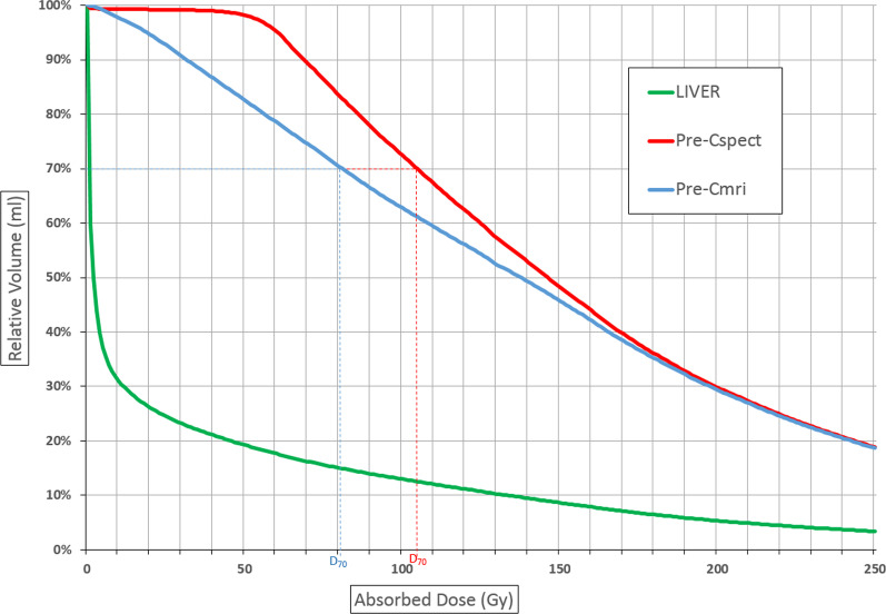
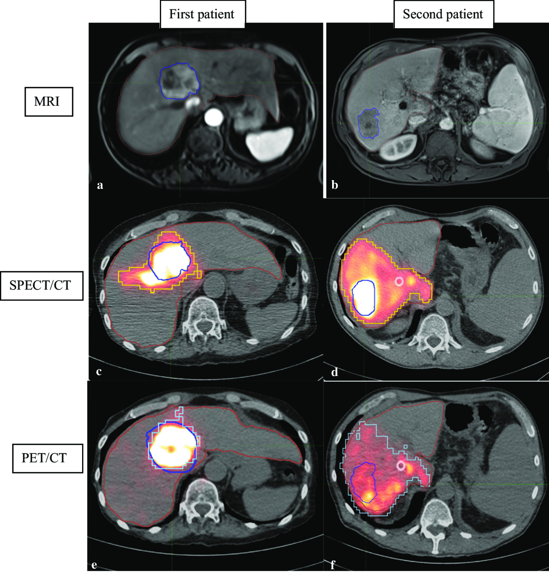
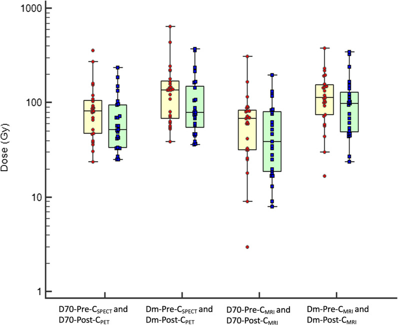
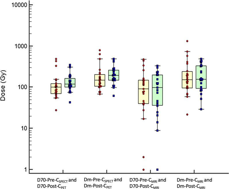
Methods: Forty-eight patients who underwent SIRT between 2012 and 2020 were retrospectively included in this study. Target volumes were delineated using two methods: MRI-based contours manually drawn by a radiologist and then registered on SPECT/CT and PET/CT via deformable registration (Pre-CMRI and Post-CMRI), 99mTc-MAA-SPECT and 90Y-microspheres-PET 10% threshold contouring (Pre-CSPECT and Post-CPET). The mean absorbed dose (Dm) and the minimal absorbed dose delivered to 70% of the tumor volume (D70) were evaluated with both contouring methods; the tumor-to-normal liver uptake ratio (TNR) was evaluated with MRI-based contours only. Tumor response was assessed using the mRECIST criteria on the follow-up MRIs.
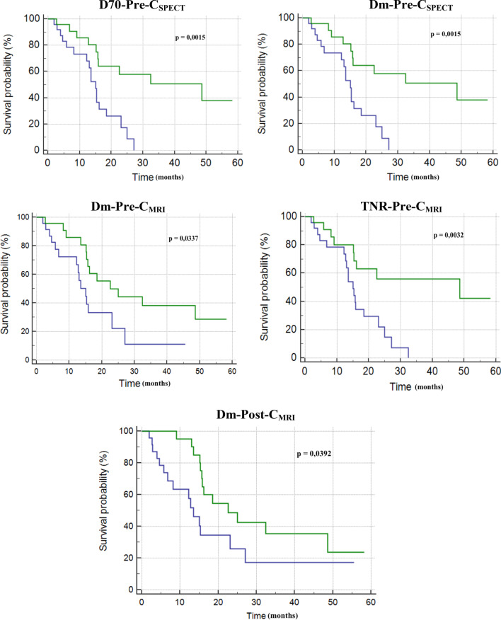
Results: No significant differences were found for Dm and TNR between pre- and post-treatment. TNR evaluated with radiologic contours (Pre-CMRI and Post-CMRI) were predictive of tumor control at 6 months on pre- and post-treatment dosimetry (OR 5.9 and 7.1, respectively; p = 0.02 and 0.01). All dose metrics determined with both methods were predictive of overall survival (OS) on pre-treatment dosimetry, but only Dm with MRI-based contours was predictive of OS on post-treatment images with a median of 23 months for patients with a supramedian Dm versus 14 months for the others (p = 0.04).
Conclusion: In advanced HCC treated with SIRT, Dm and TNR determined with radiologic contours were predictive of tumor control and OS. This study shows that a rigorous clinical workflow (radiologic contours + registration on scintigraphic images) is feasible and should be prospectively considered for improving therapeutic strategy.
Keywords: Dosimetry; Hepatocellular carcinoma; Radioembolization.
Conflict of interest statement
Romaric Loffroy, Consultant, Sirtex. All other authors declare that they have no conflict of interest.
Figures
Similar articles
-
Partition Model-Based 99mTc-MAA SPECT/CT Predictive Dosimetry Compared with 90Y TOF PET/CT Posttreatment Dosimetry in Radioembolization of Hepatocellular Carcinoma: A Quantitative Agreement Comparison.J Nucl Med. 2016 Nov;57(11):1672-1678. doi: 10.2967/jnumed.116.173104. Epub 2016 Jun 15. J Nucl Med. 2016. PMID: 27307346
-
Analysis of differences between 99mTc-MAA SPECT- and 90Y-microsphere PET-based dosimetry for hepatocellular carcinoma selective internal radiation therapy.EJNMMI Res. 2019 Jul 22;9(1):62. doi: 10.1186/s13550-019-0533-6. EJNMMI Res. 2019. PMID: 31332585 Free PMC article.
-
Semi-Quantitative Analysis of Post-Transarterial Radioembolization (90)Y Microsphere Positron Emission Tomography Combined with Computed Tomography (PET/CT) Images in Advanced Liver Malignancy: Comparison With (99m)Tc Macroaggregated Albumin (MAA) Single Photon Emission Computed Tomography (SPECT).Nucl Med Mol Imaging. 2016 Mar;50(1):63-9. doi: 10.1007/s13139-015-0366-9. Epub 2015 Sep 14. Nucl Med Mol Imaging. 2016. PMID: 26941861 Free PMC article.
-
90Y-Loaded Microsphere SIRT of HCC Patients With Portal Vein Thrombosis: High Clinical Impact of 99mTc-MAA SPECT/CT-Based Dosimetry.Semin Nucl Med. 2019 May;49(3):218-226. doi: 10.1053/j.semnuclmed.2019.01.006. Epub 2019 Feb 7. Semin Nucl Med. 2019. PMID: 30954188 Review.
-
Clinical impact of (99m)Tc-MAA SPECT/CT-based dosimetry in the radioembolization of liver malignancies with (90)Y-loaded microspheres.Eur J Nucl Med Mol Imaging. 2016 Mar;43(3):559-75. doi: 10.1007/s00259-015-3157-8. Epub 2015 Sep 4. Eur J Nucl Med Mol Imaging. 2016. PMID: 26338177 Free PMC article. Review.
Cited by
-
Segmentation-guided multi-modal registration of liver images for dose estimation in SIRT.EJNMMI Phys. 2022 Jan 25;9(1):3. doi: 10.1186/s40658-022-00432-8. EJNMMI Phys. 2022. PMID: 35076801 Free PMC article.
-
Optimization of the Clinical Effectiveness of Radioembolization in Hepatocellular Carcinoma with Dosimetry and Patient-Selection Criteria.Curr Oncol. 2022 Mar 29;29(4):2422-2434. doi: 10.3390/curroncol29040196. Curr Oncol. 2022. PMID: 35448170 Free PMC article. Review.
-
99mTc-MAA SPECT/CT imaging before 90Y-SIRT for reflecting the distribution of 90Y resin microspheres in tumors and the liver: a comparison with 90Y PET/CT verification imaging.Quant Imaging Med Surg. 2025 Jul 1;15(7):6272-6286. doi: 10.21037/qims-24-2232. Epub 2025 Jun 27. Quant Imaging Med Surg. 2025. PMID: 40727337 Free PMC article.
References
-
- Global Burden of Disease Cancer Collaboration. Fitzmaurice C, Allen C, Barber RM, Barregard L, Bhutta ZA, et al. Global, regional, and national cancer incidence, mortality, years of life lost, years lived with disability, and disability-adjusted life-years for 32 cancer groups, 1990 to 2015: a systematic analysis for the Global Burden of Disease Study. JAMA Oncol. 2017;3:524–548. doi: 10.1001/jamaoncol.2016.5688. - DOI - PMC - PubMed
-
- Loffroy R, Ronot M, Greget M, Bouvier A, Mastier C, Sengel C, et al. Short-term safety and quality of life outcomes following radioembolization in primary and secondary liver tumours: a multi-centre analysis of 200 patients in France. Cardiovasc Intervent Radiol. 2020;44:36–49. doi: 10.1007/s00270-020-02643-x. - DOI - PMC - PubMed
LinkOut - more resources
Full Text Sources
Other Literature Sources
