. 2021 Apr;35(4):e21468.
doi: 10.1096/fj.202002778R.

Targeting alveolar-specific succinate dehydrogenase A attenuates pulmonary inflammation during acute lung injury

Affiliations

Targeting alveolar-specific succinate dehydrogenase A attenuates pulmonary inflammation during acute lung injury

Christine U Vohwinkel et al. FASEB J. 2021 Apr.

Abstract

Acute lung injury (ALI) is an inflammatory lung disease, which manifests itself in patients as acute respiratory distress syndrome (ARDS). Previous studies have implicated alveolar-epithelial succinate in ALI protection. Therefore, we hypothesized that targeting alveolar succinate dehydrogenase SDH A would result in elevated succinate levels and concomitant lung protection. Wild-type (WT) mice or transgenic mice with targeted alveolar-epithelial Sdha or hypoxia-inducible transcription factor Hif1a deletion were exposed to ALI induced by mechanical ventilation. Succinate metabolism was assessed in alveolar-epithelial via mass spectrometry as well as redox measurements and evaluation of lung injury. In WT mice, ALI induced by mechanical ventilation decreased SDHA activity and increased succinate in alveolar-epithelial. In vitro, cell-permeable succinate decreased epithelial inflammation during stretch injury. Mice with inducible alveolar-epithelial Sdha deletion (Sdhaloxp/loxp SPC-CreER mice) revealed reduced lung inflammation, improved alveolar barrier function, and attenuated histologic injury. Consistent with a functional role of succinate to stabilize HIF, Sdhaloxp/loxp SPC-CreER experienced enhanced Hif1a levels during hypoxia or ALI. Conversely, Hif1aloxp/loxp SPC-CreER showed increased inflammation with ALI induced by mechanical ventilation. Finally, wild-type mice treated with intra-tracheal dimethlysuccinate were protected during ALI. These data suggest that targeting alveolar-epithelial SDHA dampens ALI via succinate-mediated stabilization of HIF1A. Translational extensions of our studies implicate succinate treatment in attenuating alveolar inflammation in patients suffering from ARDS.

Keywords: ALI; ARDS; HIF; SDHA; alveolar epithelium; inflammation; mechanical ventilation; succinate.


Conflict of interest statement

The authors declare no competing interests.

Figures
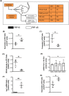
FIGURE 1
FIGURE 1
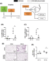
ALI induced by mechanical ventilation is associated with reduced activity of SDHA in alveolar epithelium. A, Schematic overview of ventilation experiments: 10‐ to 12‐week‐old mice were anesthetized and ventilated for 4 h with the following parameters: Pressure‐controlled ventilation (PCV), peak inspiratory pressure (PIP) 15 cm H2O in the control group and 45 cm H2O in the experimental group, PEEP 3 cm H2O, respiratory rate 80/min, and FiO2 1.0 were set equally in both groups. B and C, TCA cycle intermediates succinate and fumarate were determined with mass spectroscopy in ATII cells after mice were exposed to 15 or 45 cm H2O PIP. D, Succinate dehydrogenase (SDHA) activity was determined by calculating the succinate to fumarate ratio. mRNA expression of SDH subunits (E) and Irg‐1(F) was determined with qPCR. (G) Itaconate abundance was measured in ATII cells via mass spectroscopy. Data are represented as mean ± SD, n = 3‐4, ns‐not significant, *P <.05, **P <.01, Welch's t test in F, t = 5.993 and df = 3.09; two‐sample t test in G, t = 3.43 and df = 5. A total of 21 animals were used
FIGURE 2
FIGURE 2
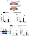
Succinate attenuates epithelial inflammation during cyclic mechanical stretch exposure. A, Schematic of in vitro cyclic mechanical stretch system. Cells were plated on a flexible membrane coated with collagen and vacuum to induce mechanical stretch to simulate injurious ventilation in vitro. B and C, mRNA expression of pro‐inflammatory cytokine IL‐6 and chemokine IL‐8 was determined in human alveolar‐epithelial cell line A549 with qPCR after 24 h of mechanical stretch and treatment with cell‐permeable 10 μM dimethlysuccinate or vehicle. D, Western blot confirmation of SDHA knockdown in A549 cells SDHA shRNA, control cells were transfected with scrambled shRNA (scrRNA). E and F, mRNA expression of IL‐6 and. IL‐8 in A549 cells with stable knockdown of SDHA (shRNA SDHA) or scrambled controls (Scr A549). Cells were exposed to 24 h of cyclic mechanical stretch or control conditions. Data are represented as mean ± SD, n = 5‐6, **P <.01, **P <.001; Welch's t test with Bonferroni adjustment
FIGURE 3
FIGURE 3
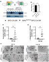
Generation and characterization of mice with alveolar epithelial deletion of Sdha. A, Generation of mice with AT II‐specific deletion of Sdha. Animals homozygous for floxed Sdha allele (Sdhaloxp/loxp) were crossed with SPC‐CreER mice to generate the Sdhaloxp/loxp SPC‐CreER line. Conditional Sdha suppression was induced by i.p. tamoxifen. Sdha suppression in AT II cells was confirmed with Western blot. B, Weights of Sdhaloxp/loxp SPC‐CreER mice compared with SPC‐CreER control mice. Both groups were age matched at 10‐12 weeks of age after having received tamoxifen for 5 days, 2 weeks prior to ventilation. C and D, Quantification of TCA cycle intermediates succinate (C) and α‐ketoglutarate (D) in ATII cells after ventilation with 45 cm2 H2O by mass spectroscopy. E, Electron microscopic images of wild‐type and Sdhaloxp/loxp SPC‐CreER animals in control (PIP 15 cm H2O) and experimental (PIP 45 cm H2O) conditions. ATII = alveolar epithelial type II, E = erythrocyte, Endo = endothelial cell, Mϕ = Macrophage, Peri = pericytes. Data are represented as mean ± SD, n = 4‐5, *P <.05, B; Welch's t test in C, t = 2.97 and df = 4; Welch's t test in D, t = 3.64 and df = 4.22. Total of 17 animals were used for panels C‐E (panel B encompasses animals from Figures 3, 4, 5)
FIGURE 4
FIGURE 4
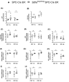
alveolar‐epithelial Sdha deletion does not affect ATP generation or ROS production. A, ATP concentrations were measured with bioluminescence assay and normalized to protein concentration of whole lung samples. B‐E, Concentration of mitochondrial ROS normalized to protein concentration of homogenized whole lung measured with EPR spin probes as nitroxide concentration (B) and enzyme activity of total superoxide dismutase‐ SOD (C), catalase‐CAT (D), and TBARS‐thiobarbituric acid reactive substances (E). F‐J, mRNA expression of ROS enzymes: SOD‐2 (F), CAT (G), HO‐1‐hem oxygenase 1 (H), GPX‐1‐gluthathione peroxidase1 (I), PRDX‐1‐peroxireducin 1 (J) from whole lung samples via qPCR. Data represented as mean ± SD, n = 4‐5, ns‐not significant, *P <.05, two‐sample t test or Welch's t test with Bonferroni adjustment. Total of 20 animals were used
FIGURE 5
FIGURE 5
Tissue‐specific alveolar‐epithelial Sdha deletion mediates lung protection during ALI: A‐E, Sdhaloxp/loxp SPC‐CreER mice or age, gender, and weight‐matched controls (SPC‐CreER) were exposed PIP of 45 cm H2O to induce lung injury or control ventilation (15 cm H2O). A and B, mRNA expression was determined from ATII cells with qPCR. C, Protein concentration was measured in bronchoalveolar lavage fluid (BALF). D, Representative images of H&E‐stained lungs from mice subjected to PIP of 15 vs 45 cm H2O. E, Cumulative lung injury score which is a combined score of cellular infiltrates, interstitial congestion and hyaline membrane formation, and hemorrhage. Data are represented as mean ± SD, n = 4‐7, ns‐not significant, *P <.05, **P <.01. Welch's t test with Bonferroni adjustment. Total of 35 animals were used
FIGURE 6
FIGURE 6
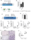
Alveolar‐epithelial Hif1a stabilization as mediator of succinate‐elicited lung protection. A and B, ATII cells were isolated from SPC‐CreER and Sdhaloxp/loxp SPC‐CreER mice exposed to 24 h of hypoxia (10% FiO2). ATII cells were lysed, and nuclear Hif1a expression was determined with Western blot and quantified. C, Generation of mice with AT II‐specific deletion of Hif1a. Animals homozygous for floxed Hif1a allele (Hif1aloxp/loxp) were crossed with SPC‐CreER mice to generate the Hif1aloxp/loxp SPC‐CreER loxp/loxp line. Conditional Hif1a suppression was induced by i.p. tamoxifen. D, Confirmation of Hif1a suppression in ATII cells with Western blot. E, Weights of Hif1a loxp/loxp SPC‐CreER mice compared with SPC‐CreER control mice. F and G, mRNA expression was determined from whole lung tissue with qPCR after mice were subjected to injurious (PIP 45 cm H2O) or control ventilation (PIP 15 cm H2O). H, Protein concentration was measured in BALF. I, Representative images of H&E‐stained lungs from mice subjected to PIP 15 cm H2O) vs PIP 45 cm H2O). J, Cumulative lung injury score which is a combined score of cellular infiltrates, interstitial congestion and hyaline membrane formation, and hemorrhage. Data are represented as mean ± SD, n = 3‐7, ns‐not significant, *P <.05, **P <.01, ****P <.0001, B and E, two‐sample t test; F‐G, unpaired, Welch's t test with Bonferroni adjustment. Total of 47 animals were used
FIGURE 7
FIGURE 7
Local delivery of dimethlysuccinate attenuates inflammation in ALI. A. Schematic overview of the experiments. Anesthetized mice received either 50 μL of vehicle (physiol. NaCl) or 10 μM dimethyl succinate i.t. 15 min prior to initiation of control (PIP 15) or injurious ventilation PIP 45); after 4 h, lungs were removed. B and C, mRNA expression was determined from whole lung tissue with qPCR after mice were subjected to ventilation with PIP 15 vs 45 cm H2O. D, Representative images of H&E‐stained lungs from mice subjected to PIP 15 vs 45 cm H2O. E, Cumulative lung injury score which is a combined score of cellular infiltrates, interstitial congestion and hyaline membrane formation, and hemorrhage. Data are represented as mean ± SD, n = 3‐4, ns‐not significant, *P <.05, Welch's t test with Bonferroni adjustment. Total of 30 animals were used, and no animal was excluded from analysis

References

    1. Williams GW, Berg NK, Reskallah A, Yuan X, Eltzschig HK. Acute respiratory distress syndrome. Anesthesiology. 2021;134(2):270‐282. - PMC - PubMed
    1. Ranieri VM, Rubenfeld GD, Thompson BT, et al. Acute respiratory distress syndrome: the Berlin Definition. JAMA. 2012;307:2526‐2533. - PubMed
    1. Eltzschig HK, Carmeliet P. Hypoxia and inflammation. N Engl J Med. 2011;364:656‐665. - PMC - PubMed
    1. Rubenfeld GD, Caldwell E, Peabody E, et al. Incidence and outcomes of acute lung injury. N Engl J Med. 2005;353:1685‐1693. - PubMed
    1. Villar J, Blanco J, Anon JM, et al. The ALIEN study: incidence and outcome of acute respiratory distress syndrome in the era of lung protective ventilation. Intensive Care Med. 2011;37:1932‐1941. - PubMed

Publication types

Substances

LinkOut - more resources