. 2021 Apr;73(2):792-827.
doi: 10.1124/pharmrev.120.000072.

Therapeutic Targeting of the Complement System: From Rare Diseases to Pandemics

Affiliations

Therapeutic Targeting of the Complement System: From Rare Diseases to Pandemics

Peter Garred et al. Pharmacol Rev. 2021 Apr.

Abstract

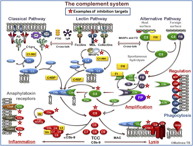
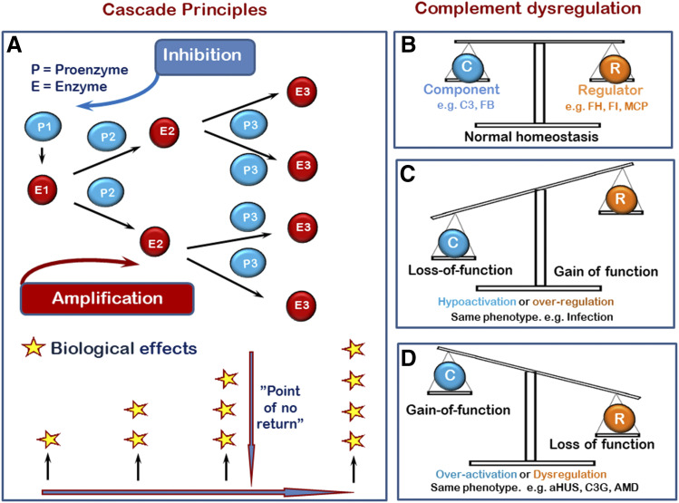
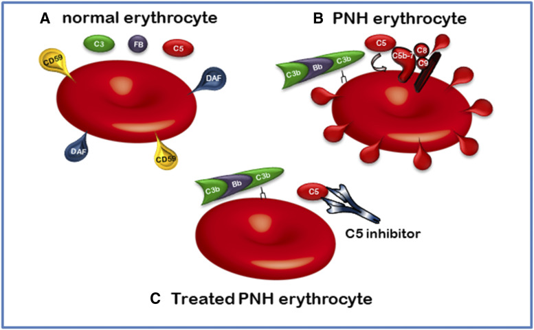
The complement system was discovered at the end of the 19th century as a heat-labile plasma component that "complemented" the antibodies in killing microbes, hence the name "complement." Complement is also part of the innate immune system, protecting the host by recognition of pathogen-associated molecular patterns. However, complement is multifunctional far beyond infectious defense. It contributes to organ development, such as sculpting neuron synapses, promoting tissue regeneration and repair, and rapidly engaging and synergizing with a number of processes, including hemostasis leading to thromboinflammation. Complement is a double-edged sword. Although it usually protects the host, it may cause tissue damage when dysregulated or overactivated, such as in the systemic inflammatory reaction seen in trauma and sepsis and severe coronavirus disease 2019 (COVID-19). Damage-associated molecular patterns generated during ischemia-reperfusion injuries (myocardial infarction, stroke, and transplant dysfunction) and in chronic neurologic and rheumatic disease activate complement, thereby increasing damaging inflammation. Despite the long list of diseases with potential for ameliorating complement modulation, only a few rare diseases are approved for clinical treatment targeting complement. Those currently being efficiently treated include paroxysmal nocturnal hemoglobinuria, atypical hemolytic-uremic syndrome, myasthenia gravis, and neuromyelitis optica spectrum disorders. Rare diseases, unfortunately, preclude robust clinical trials. The increasing evidence for complement as a pathogenetic driver in many more common diseases suggests an opportunity for future complement therapy, which, however, requires robust clinical trials; one ongoing example is COVID-19 disease. The current review aims to discuss complement in disease pathogenesis and discuss future pharmacological strategies to treat these diseases with complement-targeted therapies. SIGNIFICANCE STATEMENT: The complement system is the host's defense friend by protecting it from invading pathogens, promoting tissue repair, and maintaining homeostasis. Complement is a double-edged sword, since when dysregulated or overactivated it becomes the host's enemy, leading to tissue damage, organ failure, and, in worst case, death. A number of acute and chronic diseases are candidates for pharmacological treatment to avoid complement-dependent damage, ranging from the well established treatment for rare diseases to possible future treatment of large patient groups like the pandemic coronavirus disease 2019.


Conflict of interest statement

Conflict of Interest: T.E.M. is a member of the Scientific Advisory Board of Ra Pharma/UCB. A.J.T. is a consultant for Montis.

Figures

Fig. 1.
Fig. 1.
An overview of the complement system with focus on most relevant targets for therapeutic inhibition. The complement system acts as a recognition system and can recognized danger and be activated through three initial pathways (upper part of figure), all converging to the cleavage of C3 to generate C3a and C3b (middle part of figure). The classical pathway (CP) is typically activated by antibodies, but amyloid β fibrils and pentraxins, including CRP, serum amyloid P component (SAP), and PTX3, can activate C1. The lectin pathway (LP) is activated through recognition of carbohydrates by MBL, ficolins, and collectins. Furthermore, LP activation may be mediated through IgM antibodies (e.g., directed against damaged self antigens). Both the CP and the LP activate C4 to C4a, and C4b and C2 subsequently bind to C4b and make the C4bC2b convertase after C2 is cleaved to C2a and C2b. The alternative pathway (AP) is activated by foreign or damaged cells, as facilitated by the continuous spontaneous hydrolysis of C3. AP also has an important function in the complement system providing an amplification loop enhancing C3 activation independent of which pathway that is initially activated. This effect is enhanced due to properdin (FP), the only positive regulator in the complement system, which stabilizes the C3 convertase. Cleavage of C3 leads to formation of a C5 convertase, cleaving C5 into C5a and C5b. The anaphylatoxins C3a and C5a bind to the receptors C3aR, C5aR1, and C5aR2, leading to downstream production of proinflammatory and/or anti-inflammatory mediators (lower left part of figure). C5b initiates the formation of the TCC, which forms the membrane attack complex (MAC) if inserted into a membrane (bottom part of figure). This may lead to lysis of bacteria and cells or in sublytic doses to activation of cells. The cleavage and inactivation of C3b generate iC3b, binding to complement receptors CR3 (CD11b/CD18) and CR4 (CD11c/CD18) and facilitating phagocytosis, oxidative burst, and downstream inflammation (right part of figure). The complement system is tightly regulated by soluble inhibitors (marked in yellow), including C1-inhibitor (C1-INH), factor H (FH), factor I (FI), C4BP, carboxidase inactivation of the anaphylotoxins (AI), vitronectin (Vn), and clusterin (CLU), keeping the continuous low-grade activation in the fluid phase in check. Host cell membranes are equipped with a number of inhibitors to protect them against attack by complement (right part of figure), including MCP (CD46), CR1 (CD35), DAF (CD55) controlling C4 and C3 activation, and CD59 protecting against final assembly of the C5b-9 complex. Some selected attractive targets for therapeutic inhibition are indicated by red asterisks. Although many more could have been included, we selected C1s as a specific target from the CP, MBL, and MASP-2 from the LP, factor B, factor D, and properdin as specific for the AP and then C3 as the major component at which all three pathways converge and would be a very efficient blocker of the system. C5 is the next main candidate to block completely, as it will block the inflammatory potent C5a fragment and formation of the inflammatory and lytic C5b-9 complex or the solueble form sC5b-9. In addition, C5a can be inhibited, preserving the C5b-9 pathway, or the C5b7 can be blocked to prevent C5b-9 formation, leaving C5a open. Finally, the anaphylatoxin receptor’s axes can be blocked to prevent signaling. In particular, blocking of C5aR1 will attenuate the proinflammatory inflammation, whereas the effects of blocking C3aR and C5aR2 receptors are to be studied in more detail since they might have more anti-inflammatory effects. FB, factor B. (The figure is a modified version of one published in J Leukoc Biol (2014) 101:193–204. Barratt-Due A, Pischke SE, Nilsson PH, Espevik T, Mollnes TE. Copyright by Mollnes TE.)
Fig. 2.
Fig. 2.
Cascade principles and the need for balance and homeostasis. (A) Illustration of a cascade with the potential for amplification and thus the need for inhibition. The balance is kept under control as long as the activation occurs locally. If the activation is overwhelming, such as in systemic inflammation, the explosive response might kill the host. (B) Normal homeostasis with balance between inhibition and activation. (C) Hypoactivation due to loss of function on ordinary components or gain of function in regulators, with both leading to the same phenotype. (D) The opposite situation with gain of function in ordinary components and loss of function of regulators resulting in the same hyperactivation phenotype. FB, factor B; FH, factor H; FI, factor I.
Fig. 3.
Fig. 3.
Loss of host cell regulators leads to attack by the host’s own complement. (A) A normal erythrocyte protected from complement by DAF (CD55) and CD59. (B) An erythrocyte from a patient with PNH, which is caused by a clonal deletion in enzyme enabling GPI anchors, which is responsible for binding both DAF (CD55) and CD15 to the surface, resulting in cell lysis. (C) PNH cell from a patient treated with eculizumab, a monoclonal antibody blocking cleavage of C5. The cell is protected from C5b-9 attack. FB, factor B.
Fig. 4.
Fig. 4.
Signs and symptoms of inflammation. The classic four characteristics seen in any inflammation: calor, rubor, tumor, and dolor. Later, function loss was also added to these. Reprinted with permission from Lawrence T, Willoughby DA, and Gilroy DW (2002) Anti-inflammatory lipid mediators and insights into the resolution of inflammation. Nature Reviews Immunology 2:787–795. Printed with permission from Nat Rev Immunol.
Fig. 5.
Fig. 5.
Immunohistochemically detected deposition of complement in tissue. (A) C5aR1 antagonist (PMX205) reduces microglial cells (IBA-1) and CD68 surrounding amyloid plaques in an Alzheimer disease mouse model. Representative hippocampal images of ThioS (fAβ green), Iba1 (microglia, blue), and CD68 (lysosomal marker, red) in 15-month-old Tg2576 after treatment with the C5aR1 antagonist PMX205 (B1-B4) or untreated (A1-A4) for 12 weeks. C5aR1 antagonist treatment showed reduced microglial cells (IBA, blue) surrounding amyloid plaques (ThioS, green) and accompanied by a reduction of the lysosomal marker CD68 (red). Courtesy of Angela Gomez-Arboledas. (B) Deposition of C5b-9 in a porcine glomerulus from a case with factor-H deficiency and C3 glomerulopathy presented as dense deposit disease (kindly provided by Professor Johan Høgset Jansen). (C) C4d deposition in peritubular capillaries in a kidney undergoing acute antibody-mediated rejection (kindly provided by the Department of Pathology at Oslo University Hospital).
Fig. 6.
Fig. 6.
Immunoassay for detection of complement activation. This ELISA illustrates the first specific assay for detection of sC5b-9 based on mAbE11 produced in our laboratory in 1985 reacting specifically with a neoepitope in C9 exposed only when C9 is incorporated in the C5b-9 complex (Mollnes et al., 1985). The assay is a traditional sandwich immunoassay, and many variants exist with different detection antibodies on the top, reacting with one of the other components of the complex. An increased level of sC5b-9 indicates that the complement cascade has been activated to the very end.
Fig. 7.
Fig. 7.
Immunoassay for detection of total complement function. This assay is used to screen for complement deficiencies in each activation pathway and to evaluate the effect of treating patients with, for example, a C5 inhibitor. (A) Normal serum leading to complete assembly of the C5b-9 being similar to the membrane attack complex complex, as it is bound to the plastic surface not measuring sC5b-9. All three pathways show 100% activity. (B) Serum from a patient who is C5-deficient. The assembly will stop at the level of C5, and all three wells will have 0% activity. (C) Serum from a patient treated in excess with the mAb eculizumab blocking C5 cleavage. This will give the same phenotype as the patient who is genetically deficient. If eculizumab is discontinued it will gradually disappear, and the situation will be similar to the one presented in (A), in which the inhibitory anti-C5 mAb is not present any longer, and the activity will be 00% as before treatment started. HRP, horseradish peroxidase.

Similar articles

Cited by

References

    1. Abe T, Hosur KB, Hajishengallis E, Reis ES, Ricklin D, Lambris JD, Hajishengallis G (2012) Local complement-targeted intervention in periodontitis: proof-of-concept using a C5a receptor (CD88) antagonist. J Immunol 189:5442–5448. - PMC - PubMed
    1. Ahmad S, Pandya C, Kindelin A, Bhatia K, Chaudhary R, Dwivedi AK, Eschbacher JM, Liu Q, Waters MF, Hoda MN, et al. (2020) C3a receptor antagonist therapy is protective with or without thrombolysis in murine thromboembolic stroke. Br J Pharmacol 177:2466–2477. - PMC - PubMed
    1. Ahmad SB, Bomback AS (2020) C3 glomerulopathy: pathogenesis and treatment. Adv Chronic Kidney Dis 27:104–110. - PubMed
    1. Alawieh A, Elvington A, Tomlinson S (2015) Complement in the homeostatic and ischemic brain. Front Immunol 6:417. - PMC - PubMed
    1. Alawieh A, Langley EF, Tomlinson S (2018a) Targeted complement inhibition salvages stressed neurons and inhibits neuroinflammation after stroke in mice. Sci Transl Med 10:6459. - PMC - PubMed

Publication types

MeSH terms