Archaeal Connectase is a specific and efficient protein ligase related to proteasome β subunits
- PMID: 33688044
- PMCID: PMC7980362
- DOI: 10.1073/pnas.2017871118
Archaeal Connectase is a specific and efficient protein ligase related to proteasome β subunits
Abstract
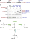
Sequence-specific protein ligations are widely used to produce customized proteins "on demand." Such chimeric, immobilized, fluorophore-conjugated or segmentally labeled proteins are generated using a range of chemical, (split) intein, split domain, or enzymatic methods. Where short ligation motifs and good chemoselectivity are required, ligase enzymes are often chosen, although they have a number of disadvantages, for example poor catalytic efficiency, low substrate specificity, and side reactions. Here, we describe a sequence-specific protein ligase with more favorable characteristics. This ligase, Connectase, is a monomeric homolog of 20S proteasome subunits in methanogenic archaea. In pulldown experiments with Methanosarcina mazei cell extract, we identify a physiological substrate in methyltransferase A (MtrA), a key enzyme of archaeal methanogenesis. Using microscale thermophoresis and X-ray crystallography, we show that only a short sequence of about 20 residues derived from MtrA and containing a highly conserved KDPGA motif is required for this high-affinity interaction. Finally, in quantitative activity assays, we demonstrate that this recognition tag can be repurposed to allow the ligation of two unrelated proteins. Connectase catalyzes such ligations at substantially higher rates, with higher yields, but without detectable side reactions when compared with a reference enzyme. It thus presents an attractive tool for the development of new methods, for example in the preparation of selectively labeled proteins for NMR, the covalent and geometrically defined attachment of proteins on surfaces for cryo-electron microscopy, or the generation of multispecific antibodies.
Keywords: methanogenic archaea; proteasome; protein ligation; sortase; transpeptidase.
Copyright © 2021 the Author(s). Published by PNAS.
Conflict of interest statement
Competing interest statement: Max Planck Innovation has filed a provisional patent on Connectase and its use for enzymatic ligation.
Figures


Similar articles
-
Structural Basis of High-Precision Protein Ligation and Its Application.J Am Chem Soc. 2025 Jan 15;147(2):1604-1611. doi: 10.1021/jacs.4c10689. Epub 2025 Jan 2. J Am Chem Soc. 2025. PMID: 39745918
-
Discovery and characterization of the first archaeal dihydromethanopterin reductase, an iron-sulfur flavoprotein from Methanosarcina mazei.J Bacteriol. 2014 Jan;196(2):203-9. doi: 10.1128/JB.00457-13. Epub 2013 Aug 30. J Bacteriol. 2014. PMID: 23995635 Free PMC article.
-
Functional and structural characterization of the Methanosarcina mazei proteasome and PAN complexes.J Struct Biol. 2006 Oct;156(1):84-92. doi: 10.1016/j.jsb.2006.03.015. Epub 2006 Apr 25. J Struct Biol. 2006. PMID: 16690322
-
Archaeal proteasomes and sampylation.Subcell Biochem. 2013;66:297-327. doi: 10.1007/978-94-007-5940-4_11. Subcell Biochem. 2013. PMID: 23479445 Free PMC article. Review.
-
Natural substrates of the proteasome and their recognition by the ubiquitin system.Curr Top Microbiol Immunol. 2002;268:137-74. doi: 10.1007/978-3-642-59414-4_6. Curr Top Microbiol Immunol. 2002. PMID: 12083004 Review.
Cited by
-
Protein degradation by human 20S proteasomes elucidates the interplay between peptide hydrolysis and splicing.Nat Commun. 2024 Feb 7;15(1):1147. doi: 10.1038/s41467-024-45339-3. Nat Commun. 2024. PMID: 38326304 Free PMC article.
-
Specific, sensitive and quantitative protein detection by in-gel fluorescence.Nat Commun. 2023 May 2;14(1):2505. doi: 10.1038/s41467-023-38147-8. Nat Commun. 2023. PMID: 37130834 Free PMC article.
-
An Automated Workflow to Address Proteome Complexity and the Large Search Space Problem in Proteomics and HLA-I Immunopeptidomics.Mol Cell Proteomics. 2025 Jul 21;24(9):101039. doi: 10.1016/j.mcpro.2025.101039. Online ahead of print. Mol Cell Proteomics. 2025. PMID: 40701202 Free PMC article.
-
Structural Basis of High-Precision Protein Ligation and Its Application.J Am Chem Soc. 2025 Jan 15;147(2):1604-1611. doi: 10.1021/jacs.4c10689. Epub 2025 Jan 2. J Am Chem Soc. 2025. PMID: 39745918
-
InvitroSPI and a large database of proteasome-generated spliced and non-spliced peptides.Sci Data. 2023 Jan 10;10(1):18. doi: 10.1038/s41597-022-01890-6. Sci Data. 2023. PMID: 36627305 Free PMC article.
References
-
- Striebel F., et al. ., Bacterial ubiquitin-like modifier Pup is deamidated and conjugated to substrates by distinct but homologous enzymes. Nat. Struct. Mol. Biol. 16, 647–651 (2009). - PubMed
-
- Lupas A., Flanagan J. M., Tamura T., Baumeister W., Self-compartmentalizing proteases. Trends Biochem. Sci. 22, 399–404 (1997). - PubMed
Publication types
MeSH terms
Substances
LinkOut - more resources
Full Text Sources
Other Literature Sources

