. 2021 Mar 16;118(11):e2020024118.
doi: 10.1073/pnas.2020024118.

The diversity and evolution of microbial dissimilatory phosphite oxidation

Affiliations

The diversity and evolution of microbial dissimilatory phosphite oxidation

Sophia D Ewens et al. Proc Natl Acad Sci U S A. .

Abstract

Phosphite is the most energetically favorable chemotrophic electron donor known, with a half-cell potential (Eo') of -650 mV for the PO43-/PO33- couple. Since the discovery of microbial dissimilatory phosphite oxidation (DPO) in 2000, the environmental distribution, evolution, and diversity of DPO microorganisms (DPOMs) have remained enigmatic, as only two species have been identified. Here, metagenomic sequencing of phosphite-enriched microbial communities enabled the genome reconstruction and metabolic characterization of 21 additional DPOMs. These DPOMs spanned six classes of bacteria, including the Negativicutes, Desulfotomaculia, Synergistia, Syntrophia, Desulfobacteria, and Desulfomonilia_A Comparing the DPO genes from the genomes of enriched organisms with over 17,000 publicly available metagenomes revealed the global existence of this metabolism in diverse anoxic environments, including wastewaters, sediments, and subsurface aquifers. Despite their newfound environmental and taxonomic diversity, metagenomic analyses suggested that the typical DPOM is a chemolithoautotroph that occupies low-oxygen environments and specializes in phosphite oxidation coupled to CO2 reduction. Phylogenetic analyses indicated that the DPO genes form a highly conserved cluster that likely has ancient origins predating the split of monoderm and diderm bacteria. By coupling microbial cultivation strategies with metagenomics, these studies highlighted the unsampled metabolic versatility latent in microbial communities. We have uncovered the unexpected prevalence, diversity, biochemical specialization, and ancient origins of a unique metabolism central to the redox cycling of phosphorus, a primary nutrient on Earth.

Keywords: CO2 fixation; Desulfotignum; Phosphitivorax; glycine reductive pathway; phosphite.


Conflict of interest statement

The authors declare no competing interest.

Figures

Fig. 1.
Fig. 1.
DPO enrichment activity. (A) Representative phosphite oxidation by the SM1 community. Temporal ion concentrations are shown for live (solid lines) or autoclaved (dashed lines) inoculum. Enrichments were amended with 10 mM phosphite at the spike point. (B) Percent change of measured ions for each enrichment community. Each row represents one community; each column displays the percent accumulation or consumption of each titled ion. Row labels are colored according to the added electron acceptor (black, CO2 only; blue, CO2 + SO42−; green, CO2 + NO3). A white dotted line denotes 50% consumption of PO33−. All percentages were calculated from concentration values prior to the first spike point. (C) Duration of ion depletion. Horizontal bars show the time frame for the metabolic activity of each measured ion. Colors correspond to B (red, PO33−; blue, SO42−; green, NO3).
Fig. 2.
Fig. 2.
Relative abundance of DPO MAGs. (A) Relative abundance of MAGs across samples. Each point represents one MAG. Color represents the presence (black) or absence (gray) of any ptx-ptd genes. (A, Top) Comparison of samples from phosphite-amended exponential phase (+Pe) with no-phosphite (−Ps) controls. (A, Bottom) Comparison of samples from phosphite-amended stationary phase (+Ps) with no-phosphite (−Ps) controls. (B) Relative abundance of MAGs across time. Each subplot represents one community, while each stacked bar represents the community composition of one sample. Colors indicate the dominant (maroon), second dominant (pink), and third dominant (yellow) DPO members, and all remaining community members (gray). Relative abundance was calculated by dividing the mean coverage of a single MAG by the sum of mean coverages for all MAGs in the respective sample.
Fig. 3.
Fig. 3.
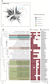
Phylogenetic trees of DPO MAGs. (A) A phylogenetic tree of bacterial genomes from the GTDB was visualized with AnnoTree (74). Nodes of the tree represent class-level taxonomy, and those nodes with DPO organisms are highlighted according to the key. (BD) Phylogenetic trees of the rpS8 marker gene showing the relationship of DPO MAGs to their closest relatives. Panels depict DPO MAGs belonging to the same phyla: (B) Firmicutes, (C) Synergistota, and (D) Desulfobacterota. The DPO MAGs from this study are bolded. Colored squares represent their dominance rank from Fig. 2B. Each close relative is annotated with its species name, accession number, and genome-source type (isolate vs. MAG), as well as its percent identity to the most closely related DPO MAG from this study. Clades are colored and labeled by taxonomic class. Internal nodes with a bootstrap support of >90% are indicated by closed circles and those with a support of >70% are indicated by open circles. (Scale bars, 0.2 change per amino acid residue.)
Fig. 4.
Fig. 4.
Metabolic model of energy conservation by Desulfomonilia_A DPOMs [adapted from Figueroa et al. (18)]. Reactions are diagramed in their hypothesized locations in relation to the inner membrane (IM) and outer membrane (OM) of a bacterial cell. Dashed lines represent mechanisms that have not been biochemically confirmed. Balanced equations are provided for phosphite oxidation and CO2 reduction to d-lactate. Dissimilatory phosphite oxidation proteins: 1) PtdC, phosphite-phosphate antiporter; 2) PtxDE-PtdFHI, putative phosphite dehydrogenase protein complex. CO2 reduction (reductive glycine pathway) proteins: 3) FdhAB/FdoGHI, formate dehydrogenase; 4) Fhs, formate:tetrahydrofolate (THF) ligase; 5) FolD, methylene-THF dehydrogenase/methenyl-THF cyclohydrolase; 6) glycine cleavage system (GcvH, lipoyl-carrier protein; GcvPAB, glycine dehydrogenase; GcvT, aminomethyltransferase; Lpd, dihydrolipoyl dehydrogenase); 7) GlyA, serine hydroxymethyltransferase; 8) SdaA/IlvA, serine dehydratase/threonine dehydratase; 9) LdhA, d-lactate dehydrogenase. Energy conversion proteins: 10) ATP synthase complex; 11) Rnf, sodium-translocating ferredoxin:NAD oxidoreductase complex; 12) NfnAB, NAD-dependent ferredoxin:NADP oxidoreductase.
Fig. 5.
Fig. 5.
Carbon and energy metabolism of DPO MAGs. Each DPO MAG was subjected to metabolic analysis via DRAM (64, 75). Within this heatmap, each cell represents a metabolic pathway (rows) for each DPO genome (columns). The number of genes for a given pathway is described by percent completion ranging from 0% (white) to 100% (brown). Pathways are organized into modules related to carbon metabolism, electron transport chain complexes, and other enzymes referenced in the text. Organisms are annotated with their taxonomic class.
Fig. 6.
Fig. 6.
CO2-dependent DPO activity. Growth and phosphite concentrations were temporally monitored in the presence and absence of CO2 for the SV3 community. Autoclaved controls showed no activity. Error bars represent SD of triplicate cultures.
Fig. 7.
Fig. 7.
Phylogenetic trees of the phosphite dehydrogenase PtxD. (A) The PtxDs from IMG/M metagenomes and DPO MAGs were aligned with proteins from the 2-hydroxyacid dehydrogenase family (Pfam PF00389; set representative proteomes to 15%). Protein subfamilies were assigned based on Matelska et al. (41). An arrow indicates the location of PtxD proteins that are associated with DPO-PtdC but clade with assimilatory phosphite oxidation PtxD (APO). (Scale bar, 0.5 change per amino acid residue.) (B) Refined tree of all PtxDs within the DPO-PtxD clade. PtxDs from the IMG/M are in light black font and labeled with their source environment and scaffold ID. PtxDs from our enriched DPO MAGs are bolded and labeled with their bacterial host name. PtxDs that belong to a binned organism are highlighted based on their taxonomic class. Published organisms with validated DPO activity are in red font. Only genes adhering to the IMG/M data usage policy are shown. Internal nodes with a bootstrap support of >70% are indicated by closed circles and those with a support of >50% are indicated by open circles. (Scale bar, 0.3 change per amino acid residue.) (C) The presence (maroon) or absence (light pink) of ptx-ptd genes in each genome was determined using custom pHMMs. Genes that were absent from a DPO MAG but present in the assembly are in gray, where phylogeny, tanglegrams, and synteny were collectively used to predict the most likely host. (D) Horizontal gray bars display the size (bp) of the contig on which each PtxD was found and are in logarithmic scale to visualize the full range of contig lengths. The black dotted line indicates the minimum length for all seven ptx-ptd genes to be present, based on FiPS-3 sequences (7,137 bp). Asterisks signify contigs that were binned.

References

    1. Pasek M. A., Sampson J. M., Atlas Z., Redox chemistry in the phosphorus biogeochemical cycle. Proc. Natl. Acad. Sci. U.S.A. 111, 15468–15473 (2014). - PMC - PubMed
    1. Figueroa I. A., Coates J. D., Microbial phosphite oxidation and its potential role in the global phosphorus and carbon cycles. Adv. Appl. Microbiol. 98, 93–117 (2017). - PubMed
    1. Pasek M. A., Rethinking early Earth phosphorus geochemistry. Proc. Natl. Acad. Sci. U.S.A. 105, 853–858 (2008). - PMC - PubMed
    1. Herschy B., et al. ., Archean phosphorus liberation induced by iron redox geochemistry. Nat. Commun. 9, 1346 (2018). - PMC - PubMed
    1. Britvin S. N., Murashko M. N., Vapnik Y., Polekhovsky Y. S., Krivovichev S. V., Earth’s phosphides in Levant and insights into the source of Archean prebiotic phosphorus. Sci. Rep. 5, 8355 (2015). - PMC - PubMed

Publication types