. 2021 Apr 1;148(7):dev193565.
doi: 10.1242/dev.193565. Epub 2021 Apr 15.

A single-plasmid approach for genome editing coupled with long-term lineage analysis in chick embryos

Affiliations

A single-plasmid approach for genome editing coupled with long-term lineage analysis in chick embryos

Shashank Gandhi et al. Development. .

Erratum in

Abstract

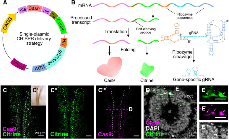
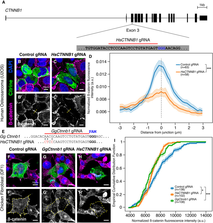
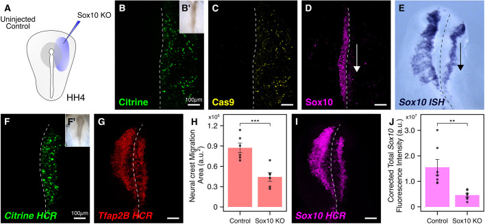
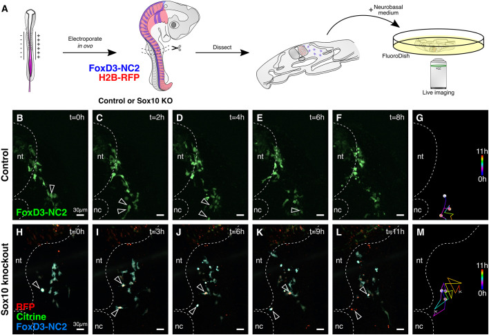
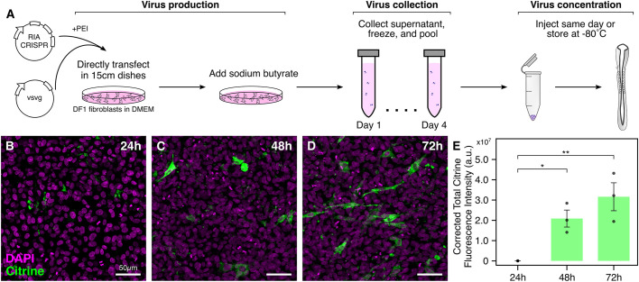
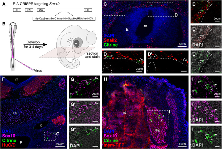
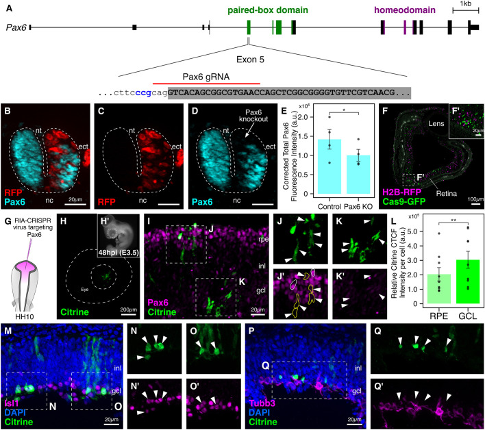
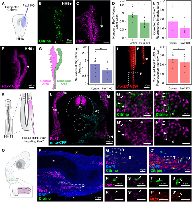
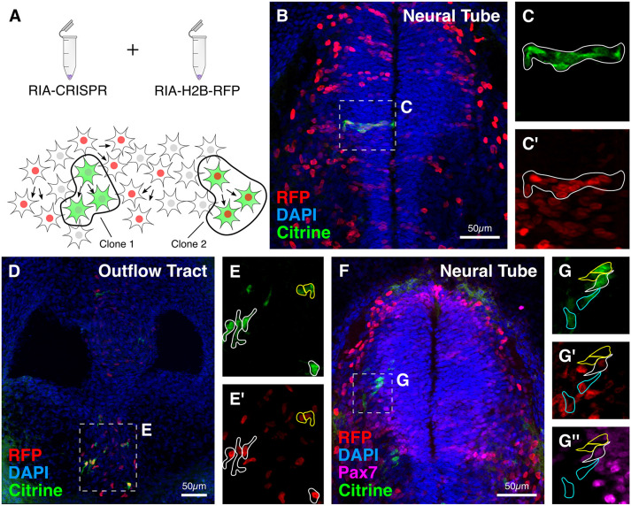
An important strategy for establishing mechanisms of gene function during development is through mutation of individual genes and analysis of subsequent effects on cell behavior. Here, we present a single-plasmid approach for genome editing in chick embryos to study experimentally perturbed cells in an otherwise normal embryonic environment. To achieve this, we have engineered a plasmid that encodes Cas9 protein, gene-specific guide RNA (gRNA), and a fluorescent marker within the same construct. Using transfection- and electroporation-based approaches, we show that this construct can be used to perturb gene function in early embryos as well as human cell lines. Importantly, insertion of this cistronic construct into replication-incompetent avian retroviruses allowed us to couple gene knockouts with long-term lineage analysis. We demonstrate the application of our newly engineered constructs and viruses by perturbing β-catenin in vitro and Sox10, Pax6 and Pax7 in the neural crest, retina, and neural tube and segmental plate in vivo, respectively. Together, this approach enables genes of interest to be knocked out in identifiable cells in living embryos and can be broadly applied to numerous genes in different embryonic tissues.

Keywords: CRISPR-Cas9; Chick embryology; Clonal analysis; Genome editing; Migration; Neural crest; Retroviruses.


Conflict of interest statement

Competing interests The authors declare no competing or financial interests.

Figures

Fig. 1.
Fig. 1.
Optimizing delivery of CRISPR components in chick embryos. (A) A single-plasmid engineered vector for optimal delivery of Cas9, gene-specific gRNA, and the fluorescent protein Citrine in transfected cells. (B) After transfection, the cistronic transcript is cleaved at the HH and HDV ribozyme sites, thereby releasing the gene-specific gRNA. Following translation of the remaining transcript, Cas9 and Citrine are separated through self-cleavage of the 2A peptide. (C-C‴) Embryos electroporated with the plasmid depicted in A at HH9 (C′) and stained with antibodies against Citrine (C″) and Cas9 (C‴) to verify cellular localization. Dashed outline delineates the neural tube. (D) A transverse cross-section through the embryo shown in C, at the indicated level, with DAPI-stained nuclei. (E-E″) Enlarged views of the boxed area in D, showing that Citrine was enriched in the cytoplasm (E, arrowheads), whereas Cas9, in the absence of a gene-specific gRNA, was restricted to the nucleolus (E′, arrowheads) within the DAPI-stained nuclei (E″). ect, ectoderm; nc, notochord; nt, neural tube.
Fig. 2.
Fig. 2.
A single CRISPR plasmid efficiently perturbs function across multiple species. (A) The genomic locus for CTNNB1 in the human genome. A gRNA protospacer targeting the third exon was designed. (B-C′) Representative images from human epithelial cells following transfection with the single CRISPR plasmid harboring control gRNA (B,B′) and human CTNNB1-targeted gRNA (C,C′). (D) Mean fluorescence intensity from line scan analysis (dashed lines in B′,C′) shows that junctions between transfected cells display reduced β-catenin immunoreactivity following CTNNB1 knockout [Kolmogorov–Smirnov test, ***P<0.001, n=63 (control gRNA) and n=58 (CTNNB1 gRNA)]. (E) Sequence alignment surrounding the Ctnnb1 gRNA protospacer between human and chicken. Red characters within the HsCTNNB1 gRNA protospacer indicate non-conserved nucleotides (asterisks). (F-H′) Representative images from chicken fibroblast cell culture following transfection with the single CRISPR plasmid harboring control gRNA (F,F′), chicken Ctnnb1-targeted gRNA (G,G′) and human CTNNB1-targeted gRNA (H,H′). Dashed yellow line in F′-H′ represents the cell boundary that was used to calculate cell fluorescence. (I) Empirical cumulative distribution frequency analysis indicates that gRNAs targeting human or chicken Ctnnb1 cause significant reduction of β-catenin immunolabeling within chicken fibroblasts compared with non-binding control gRNA transfections [Kruskal–Wallis test, *P<0.05, **P<0.01, n=138 (control gRNA), n=102 (HsCTNNB1 gRNA), n=108 (GgCtnnb1 gRNA)]. a.u., arbitrary units. Shaded areas in D and I represent s.e.m.
Fig. 3.
Fig. 3.
Electroporation-mediated early knockout of Sox10 using a single CRISPR plasmid. (A) Electroporation strategy to target Sox10 in the neural crest. The right side of gastrula-stage HH4 embryos was electroporated with a single CRISPR plasmid containing validated gRNA protospacer. (B-C) Embryos electroporated with the knockout construct developed to HH9 (B′) and immunostained for the expression of Citrine (B) and Cas9 (C). (D) The single-plasmid approach efficiently reduced Sox10 protein levels in emigrating cranial neural crest cells at HH9. (E) Chromogenic in situ hybridization (ISH) revealed a notable reduction in Sox10 mRNA levels in migrating cranial neural crest cells on the knockout side at HH9+. (F-J) Knockout embryos (F′) were processed for in situ HCR against Citrine (F), Tfap2B (G) and Sox10 (I). Quantification of migration area (H) and Sox10 fluorescence intensity (J) showed significant reduction in total area occupied by cranial neural crest cells (H; paired Student's t-test, ***P<0.001, n=6), and Sox10 mRNA levels (J; paired Student's t-test, **P<0.01, n=7) following single-CRISPR-plasmid-mediated Sox10 knockout. a.u., arbitrary units. Error bars represent s.e.m. Dashed line in image panels marks the midline.
Fig. 4.
Fig. 4.
Live imaging application of the single CRISPR plasmid. (A) Strategy for processing control and mutant embryos for live imaging. Neural crest cells were labeled using the FoxD3 enhancer NC2; knockout embryos were also electroporated with the single CRISPR plasmid targeting Sox10 and H2B-RFP as a transfection marker. (B-F) In control embryos, trunk neural crest cells divide (B,C, arrowheads) and migrate properly (D-F), with the daughter cells leaving the imaging frame/plane in E and F. (G) Tracking migration of the cells highlighted in C reveals proper trunk neural crest migration. (H-L) When Sox10 was knocked out before neural crest emigration, mutant cells still divided (H,I, arrowheads), but migrated in a circle (J,K) before undergoing apoptosis (L). (M) Cell tracking of mutant cells shows aberrant migration. Dashed line represents neural tube (nt) and notochord (nc).
Fig. 5.
Fig. 5.
Incorporating the single CRISPR plasmid into RIA retroviruses. (A) Illustration of optimized protocol to synthesize high-titer avian retroviruses that contain large inserts. Viral particles are produced in 15-cm plates supplemented with sodium butyrate. Supernatant can be pooled and stored at −80°C for several months, and concentrated virus can be injected the same day or stored at −80°C for a few weeks. (B-E) Chicken DF-1 fibroblasts were infected with RIA-CRISPR retrovirus and collected 24 h (B), 48 h (C) and 72 h (D) post-infection. Citrine expression was first observed at the 48 h time point, as validated by the quantification of Citrine CTCF intensity (E; ANOVA followed by Tukey's HSD, **P<0.01, *P<0.05, n=3 biological replicates). Error bars represent s.e.m.
Fig. 6.
Fig. 6.
RIA retrovirus-mediated knockout of Sox10 in neural crest derivatives. (A) Schematic for cloning the Sox10-RIA-CRISPR virus plasmid. (B) Sox10-RIA-CRISPR retrovirus was injected into the neural tube lumen at HH10. At HH25, embryos were fixed, cryosectioned and immunostained. (C) Knockout of Sox10 following emigration from the neural tube does not affect survival but rather causes downregulation of Snail2, a neural crest specification marker. (D-E″) Early (D,D′) and late (E-E″) migrating neural crest cells labeled with Citrine (D′,E″) do not express Snail2 (arrowheads). (F-G″) Transverse section through an injected embryo (F) shows labeled migrating trunk neural crest cells mutant for Sox10 expression close to the sympathetic ganglion (G,G′), with DAPI-labeled nuclei (G″). (H) Labeled cells were also observed in the dorsal root ganglion. (I-I″) Although most Citrine-positive cells were Sox10-negative (unfilled arrowheads), Sox10 protein was detected in a couple of labeled cells (filled arrowheads). drg, dorsal root ganglion; ic, internal carotid; nc, notochord; nt, neural tube; p, pharynx. Dashed line delineates the neural tube in D-E″, and the boundary of the dorsal root ganglion in I-I″.
Fig. 7.
Fig. 7.
Targeting Pax6 in the developing chick retina using RIA-CRISPR retrovirus. (A) The genomic locus for Pax6 in the chick with paired-box (green) and homeodomain (purple) indicated. Of gRNAs tested, the one targeting the splice acceptor site of exon 5 was most efficient. (B-E) The right side of gastrula-stage embryos was electroporated with constructs encoding Cas9, RFP and Pax6 gRNA. Transfected cells (C) lacked expression of Pax6 (D), as seen in the overlay image (B). Dashed line delineates the neural tube. Cross-sections through embryos electroporated with the single CRISPR plasmid targeting Pax6 were used to quantify fluorescence intensity of Pax6 in the neural tube; results reveal a significant difference in Pax6 expression between the knockout and control sides (E; *P<0.05, paired Student's t-test, n=4 sections from 2 representative embryos). (F,F′) Section through the eye at embryonic day 4 (F) injected with RIA-Cas9-GFP and RIA-H2B-RFP retroviruses. Labeled cells were evenly distributed (F′) through all layers of the developing retina. (G) The injection strategy for targeting Pax6 in retinal progenitor cell precursors residing in the optic vesicle. (H,H′) Citrine expression (H) in the developing eye (H′) was first observed 48 h post-infection. Dashed lines delineate the developing eye. (I-K′) Citrine-labeled cells (I,J,K) in both the retinal pigmented epithelium and the ganglion cell layers were negative for Pax6 expression (J′,K′, arrowheads). Intensity of Citrine expression together with endogenous Pax6 levels allowed identification of two clones in the retinal pigmented epithelium layer (J′): outlined in white – high Citrine, high Pax6; outlined in yellow – low Citrine, no Pax6. (L) Relative Citrine CTCF intensity per labeled cell was significantly higher in the ganglion cell layer (GCL) compared with the retinal pigmented epithelium (RPE) (*P<0.01, paired Student's t-test, n=9). (M-O′) 77.08±5.5% of Citrine-labeled cells (M,N,O) were biased towards an amacrine cell fate based on expression of Islet1 (N′,O′, arrowheads). Several migratory cells were observed in the inner nuclear layer. (P-Q′) 80.24±3.1% of Citrine-labeled cells (P,Q) in the ganglion cell layer failed to turn on Tubb3, a marker for differentiated mature neurons (Q′, arrowheads). ect, ectoderm; gcl, ganglion cell layer; inl, inner nuclear layer; nc, notochord; nt, neural tube; rpe, retinal pigmented epithelium. Error bars represent s.e.m.
Fig. 8.
Fig. 8.
Targeting Pax7 in the dermomyotome and hindlimb using RIA-CRISPR retrovirus. (A) Electroporation strategy for knocking out Pax7 in the neural crest. The right side of gastrula-stage embryos was electroporated with the single CRISPR plasmid. (B) Embryos developed to HH9+ displayed robust expression of Citrine on the right side. Dashed lines mark the midline. (C-E) The engineered single plasmid efficiently knocked out Pax7 (C) on the electroporated side. The number of neural crest cells (D; *P<0.05, paired Student's t-test, n=5) and Pax7 fluorescence intensity (E; *P<0.05, paired Student's t-test, n=5) were significantly reduced on the knockout compared with control side. (F-H) In situ HCR showed that Pax7 mRNA levels were also downregulated on the knockout side. The arrow reflects downregulation on the right side. The boundary of migration area (G) occupied by the cranial neural crest cells in F was identified using the position of the lateral-most leader cell for both control (magenta) and knockout (green) sides. Quantification of this difference revealed a significant reduction in neural crest migration on the knockout side (H; **P<0.01, paired Student's t-test, n=9). (I-J) In the absence of Pax7, the expression of the neural crest specifier gene FoxD3 is severely downregulated (I), especially in the hindbrain, where the effect of the CRISPR plasmid is most penetrant (I′). Quantification of the FoxD3 fluorescence intensity revealed significant difference between knockout and control sides (J; *P<0.05, paired Student's t-test, n=4). (K) Injection strategy for targeting Pax7 in the presomitic mesoderm of HH11 embryos using the Pax7-RIA-CRISPR retrovirus. (L) Transverse section through embryos developed to HH25 shows positive labeling with tracer virus (mito-CFP) on the injected side. (M-N′) Citrine+ cells in the dorsal (M,M′) and ventral (N,N′) aspects of the dermomyotome were Pax7-negative (arrowheads). (O) In infected embryos developed to embryonic day 6, the limb was dissected and sectioned along the coronal plane, shown in magenta. (P) Several Citrine+ mutant cells were observed in the dorsal and ventral muscle mass. (Q-U′) Pax7-mutant cells (Q,S,S′) robustly turn on the skeletal muscle marker MF20 (Q′,T-U′). Several Citrine+/MF20 cells that failed to turn on Pax7 (R,R′, arrowheads) were observed close to ventral and dorsal muscle masses, perhaps corresponding to muscle satellite cell precursors. Q and Q′ are adjacent sections that captured the same cell. do, dorsal; d, dermatome; dm, dermomyotome; dml, dorsomedial lip; drg, dorsal root ganglion; lc, limb cartilage; m, myotome; nc, notochord; nt, neural tube; v, ventral. Error bars represent s.e.m.
Fig. 9.
Fig. 9.
Proof-of-principle experiment to show application of RIA-CRISPR retroviruses for clonal analysis. (A) Experimental design for performing clonal analysis using RIA-CRISPR retroviruses. Clones were identified as single-infected cells with shared fluorescence intensity for Citrine, or double-infected cells with shared fluorescence intensity for Citrine and RFP. (B-E′) Examples of clonally related cells observed in the medial neural tube (B) and developing outflow tract (D). In the neural tube, clones (outlined) were horizontally distributed (C,C′), whereas in the outflow tract, clones were distributed evenly within the aorticopulmonary septum (E,E′). (F) In a representative embryo, double labeling with Citrine and RFP, together with intensity of Citrine and RFP and expression levels of Pax7, indicate clonal relationships. (G-G″) This allowed identification of three distinct clones: outlined in white – high Citrine (G), low RFP (G′), low Pax7 (G″); outlined in cyan – low Citrine (G), high RFP (G′), no Pax7 (G″); outlined in yellow – high Citrine (G), no RFP (G′), no Pax7 (G″).

Comment in

Similar articles

Cited by

References

    1. Andreason, G. L. and Evans, G. A. (1988). Introduction and expression of DNA molecules in eukaryotic cells by electroporation. BioTechniques 6, 650-660. - PubMed
    1. Bernsen, J. (1986). Dynamic thresholding of grey-level images. In Proceedings of the Eighth International Conference on Pattern Recognition, pp. 1251-1255. IEEE Computer Society Press.
    1. Britsch, S., Goerich, D. E., Riethmacher, D., Peirano, R. I., Rossner, M., Nave, K. A., Birchmeier, C. and Wegner, M. (2001). The transcription factor Sox10 is a key regulator of peripheral glial development. Genes Dev. 15, 66-78. 10.1101/gad.186601 - DOI - PMC - PubMed
    1. Buckingham, M., Bajard, L., Chang, T., Daubas, P., Hadchouel, J., Meilhac, S., Montarras, D., Rocancourt, D. and Relaix, F. (2003). The formation of skeletal muscle: from somite to limb. J. Anat. 202, 59-68. 10.1046/j.1469-7580.2003.00139.x - DOI - PMC - PubMed
    1. Canto-Soler, M. V. and Adler, R. (2006). Optic cup and lens development requires Pax6 expression in the early optic vesicle during a narrow time window. Dev. Biol. 294, 119-132. 10.1016/j.ydbio.2006.02.033 - DOI - PubMed

Publication types

MeSH terms

Substances