. 2021 Mar 31;148(7):dev191197.
doi: 10.1242/dev.191197.

An early global role for Axin is required for correct patterning of the anterior-posterior axis in the sea urchin embryo

Affiliations

An early global role for Axin is required for correct patterning of the anterior-posterior axis in the sea urchin embryo

Hongyan Sun et al. Development. .

Abstract

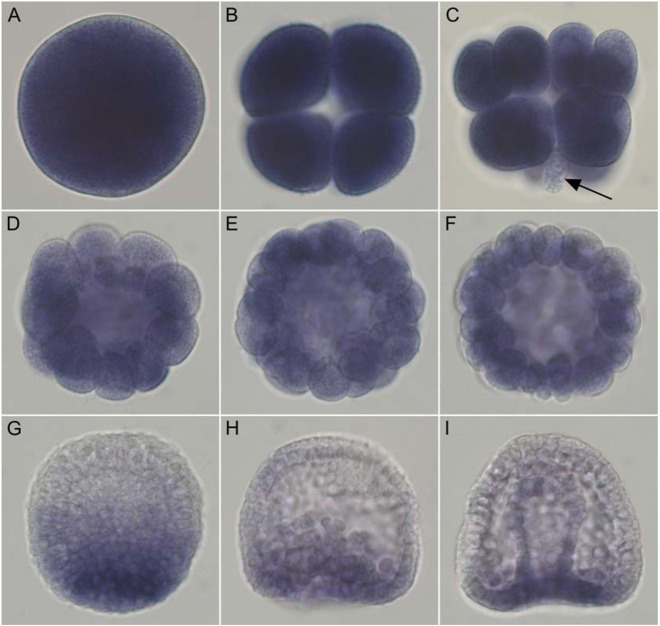
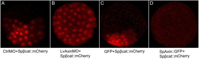
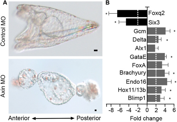
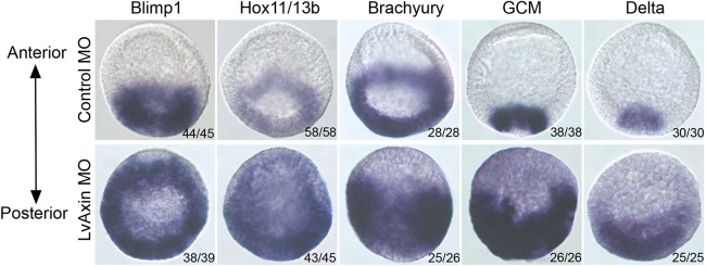
Activation of Wnt/β-catenin (cWnt) signaling at the future posterior end of early bilaterian embryos is a highly conserved mechanism for establishing the anterior-posterior (AP) axis. Moreover, inhibition of cWnt at the anterior end is required for development of anterior structures in many deuterostome taxa. This phenomenon, which occurs around the time of gastrulation, has been fairly well characterized, but the significance of intracellular inhibition of cWnt signaling in cleavage-stage deuterostome embryos for normal AP patterning is less well understood. To investigate this process in an invertebrate deuterostome, we defined Axin function in early sea urchin embryos. Axin is ubiquitously expressed at relatively high levels in early embryos and functional analysis revealed that Axin suppresses posterior cell fates in anterior blastomeres by blocking ectopic cWnt activation in these cells. Structure-function analysis of sea urchin Axin demonstrated that only its GSK-3β-binding domain is required for cWnt inhibition. These observations and results in other deuterostomes suggest that Axin plays a crucial conserved role in embryonic AP patterning by preventing cWnt activation in multipotent early blastomeres, thus protecting them from assuming ectopic cell fates.

Keywords: Animal-vegetal axis; Anterior-posterior axis; Axin; Endomesoderm; Sea urchin; Wnt signaling.


Conflict of interest statement

Competing interestsThe authors declare no competing or financial interests. Author contributionsConceptualization: H.S., C.J.P., A.H.W.; Methodology: H.S., C.J.P., H.F., A.H.W.; Validation: H.S., C.J.P., L.W., A.H.W.; Formal analysis: H.S., C.J.P., L.W., H.F., A.H.W.; Investigation: H.S., C.J.P., L.W., A.H.W.; Resources: A.H.W.; Data curation: H.S., C.J.P., L.W., H.F., A.H.W.; Writing - original draft: H.S., C.J.P., A.H.W.; Writing - review & editing: H.S., A.H.W.; Visualization: H.S.; Supervision: A.H.W.; Project administration: A.H.W.; Funding acquisition: A.H.W.

Figures

Fig. 1.
Fig. 1.
SpAxin mRNA is expressed maternally and dynamically throughout embryogenesis. Images show the spatial distribution of Axin mRNA detected by whole-mount RNA in situ hybridization in S. purpuratus eggs and embryos. (A-F) Axin is ubiquitously expressed in the egg (A), and at the 8-cell stage (B), the 16-cell stage (C), 32-cell stage (D), 60-cell stage (E) and 120-cell stage (F). At the 16-cell stage, Axin displays lower expression in the vegetal micromeres (arrow in C). (G) At the hatching blastula stage, Axin mRNA is downregulated in anterior blastomeres. (H,I) By the early gastrula stage, Axin expression is restricted to the vegetal plate (H); by the late gastrula stage, expression is downregulated in the archenteron (I).
Fig. 2.
Fig. 2.
Axin levels affect nuclearization of β-catenin in all blastomeres in sea urchin embryos. (A) Embryos co-injected with control MO and Spβ-catenin::mCherry mRNA. β-Catenin::mCherry nuclearization is seen in posterior blastomeres, as expected. (B) Embryos co-injected with Axin MO and Spβ-catenin::mCherry mRNA. Nuclear β-catenin::mCherry is seen throughout the embryo. (C) Embryos co-injected with GFP and Spβ-catenin::mCherry mRNA. Nuclear β-catenin::mCherry was seen enriched at the posterior pole. (D) When Axin and Spβ-catenin::mCherry mRNA were co-injected, no nuclear β-catenin::mCherry was observed. For each case, we observed over 100 embryos and collected images from at least seven embryos. Experiments were carried out in L. variegatus.
Fig. 3.
Fig. 3.
Knockdown of Axin posteriorizes sea urchin embryos. (A) Top panel shows a control pluteus-stage larva and the bottom panel shows an Axin-knockdown embryo at the same stage. (B) Gene expression in control and Axin-knockdown embryos at the hatching blastula stage analyzed using qPCR. The bar graph shows the fold change in expression of each gene between Axin MO- and control MO-injected embryos. Blimp1, Hox11/13b, Endo16, Brachyury, FoxA, GataE, Alx1, Delta and GCM are endomesoderm gene markers; Foxq2 and Six3 are ANE markers. qPCR experiments were replicated three times with three technical replicates for each experiment. Dashed line indicates a two-fold change. Data are mean±s.e.m. *P<0.05. Scale bars: 10 µm. Experiments were carried out in S. purpuratus.
Fig. 4.
Fig. 4.
Axin knockdown leads to ectopic expression of endomesoderm genes in anterior blastomeres. Expression of selected endomesodermal gene markers in control (top) and Axin-knockdown (bottom) L. variegatus embryos was detected using whole-mount in situ hybridization. The number of embryos showing the expression pattern shown in the figures is indicated.
Fig. 5.
Fig. 5.
Axin knockdown induces endoderm formation in isolated anterior blastomeres. (A) Protocol for isolating animal halves following morpholino injection. (B) Control pluteus larva. (B′) Axin-knockdown embryo at same stage as in B. (C,C′) Embryoids from control animal half (C) and Axin-knockdown animal half (C′). (D,D′) Endo1 expression in isolated animal halves injected with control or Axin morpholinos. Magenta, Endo1; blue, DAPI; green, phalloidin. Scale bars: 10 µm. Experiments were carried out in L. variegatus.
Fig. 6.
Fig. 6.
Axin overexpression anteriorizes sea urchin embryos. (A,C) Control embryos. (B,D) Axin-overexpressing embryos. (E) Gene expression in Axin-overexpressing embryos. The expression of selected gene markers in Axin and GFP mRNA-injected embryos at the hatching blastula stage was compared using qPCR. The bar graph shows the fold change of each gene between Axin mRNA-injected and GFP mRNA-injected control embryos. Blimp1, Hox11/13b, Endo16, Brachyury, FoxA, GataE, Alx1, Delta and GCM are endomesoderm gene markers; Foxq2 and Six3 are ANE markers. qPCR experiments were replicated with three separate batches of embryos with three technical replicates in each experiment. Dashed line indicates a twofold change in gene expression. Scale bars: 10 µm. For each experiment, 200-300 embryos were injected for each construct, and more than 95% of embryos had the same morphology as shown in the figures. *P<0.05. Experiments were carried out in S. purpuratus.
Fig. 7.
Fig. 7.
The GSK-3β-binding domain of Axin is required for rescue of Axin-knockdown embryos. (A,A′) Control embryos. (B,B′) Axin-knockdown embryos. (C,C′) Axin MO and GFP mRNA co-injected embryos. These embryos are posteriorized similar to those injected with Axin MO only (B,B′). (D,D′) Axin MO and Axin mRNA co-injected embryos. These embryos are indistinguishable from control MO-injected embryos (A,A′). (E,E′) Axin MO and Axin ΔDAX mRNA, and (F,F′) Axin MO and Axin Δβcat mRNA co-injected embryos. The Axin-knockdown phenotype is rescued in these embryos. (G,G′) Axin MO and Axin ΔRGS mRNA co-injected embryos. The Axin-knockdown phenotype is rescued in these embryos but when controls are at the pluteus stage, these embryos consistently have defects in the formation of the oral hood. Compare G′ with A′,D′,E′,F′. (H,H′) Axin MO and Axin ΔGID mRNA co-injected embryos. The Axin-knockdown phenotype is not rescued in these embryos. Each experiment was repeated three times. The numbers shown in each panel represent the number of embryos showing the phenotype shown in the panel out of the total number counted in an experiment. Scale bars: 10 µm. Arrowheads indicate the oral hood. Experiments were carried out in L. variegatus.
Fig. 8.
Fig. 8.
Structure-function analysis of Axin function in regulating anterior-posterior axis patterning. (A) The morphology of gastrula stage sea urchin embryos overexpressing full-length Axin and each of the single domain deletion constructs. Overexpression of full-length Axin, Axin Δβcat and Axin ΔDIX constructs by mRNA injection into zygotes blocked gastrulation, downregulated endomesoderm gene expression and increased ANE gene expression. Overexpression of Axin ΔRGS led to embryos that did not gastrulate, but qPCR analysis showed relatively normal gene expression, indicating that they were not anteriorized. Overexpression of Axin ΔGID had no effect on embryo development, indicating that this domain is required for the anteriorizing effect on sea urchin embryos when overexpressed. (B) Gene expression in hatching blastula stage sea urchin embryos overexpressing full-length Axin and each of the single domain deletion constructs. The y-axis shows fold change in gene expression between embryos expressing Axin constructs and embryos expressing GFP. Dashed line indicates a twofold change. Data are mean±s.e.m. *P<0.05. A scale break is used on the y-axis (−20 to −80) to adjust for the level of Hox11/13b fold change in the Axin ΔDIX-overexpressing embryos. The reason for this steep downregulation of Hox11/13b expression in Axin ΔDIX-overexpressing embryos is not known. Experiments were carried out in S. purpuratus.
Fig. 9.
Fig. 9.
Overexpression of the GID domain of Axin posteriorizes sea urchin embryos. (Aa-c) Control embryos. (Aa′-c′) Axin GID::GFP mRNA-injected embryos. By the time the control animals are at the gastrula and pluteus stages, Axin GID::GFP-expressing animals have a severely posteriorized phenotype. Scale bars: 10 µm. (B) Gene expression in Axin GID::GFP-overexpressing hatching blastula stage embryos was assayed using qPCR. qPCR experiments were replicated three times with three technical replicates for each experiment. Dashed line indicates a twofold change. Data are mean±s.e.m. *P<0.05. Experiments were carried out in S. purpuratus.

Similar articles

Cited by

References

    1. Aberle, H., Bauer, A., Stappert, J., Kispert, A. and Kemler, R. (1997). β-catenin is a target for the ubiquitin-proteasome pathway. EMBO J. 16, 3797-3804. 10.1093/emboj/16.13.3797 - DOI - PMC - PubMed
    1. Ansari, S., Troelenberg, N., Dao, V. A., Richter, T., Bucher, G. and Klingler, M. (2018). Double abdomen in a short-germ insect: Zygotic control of axis formation revealed in the beetle Tribolium castaneum. Proc. Natl. Acad. Sci. USA 115, 1819-1824. 10.1073/pnas.1716512115 - DOI - PMC - PubMed
    1. Beane, W. S., Gross, J. M. and McClay, D. R. (2006). RhoA regulates initiation of invagination, but not convergent extension, during sea urchin gastrulation. Dev. Biol. 292, 213-225. 10.1016/j.ydbio.2005.12.031 - DOI - PubMed
    1. Bince, J. M. and Wikramanayake, A. H. (2008). Functional analysis of Wnt signaling in the early sea urchin embryo using mRNA microinjection. Methods Mol. Biol. 469, 213-222. 10.1007/978-1-60327-469-2_16 - DOI - PubMed
    1. Bince, J. M., Peng, C. F. and Wikramanayake, A. H. (2008). Detecting expression patterns of Wnt pathway components in sea urchin embryos. Methods Mol. Biol. 469, 201-211. 10.1007/978-1-60327-469-2_15 - DOI - PubMed

Publication types

MeSH terms

LinkOut - more resources