. 2015 May;18(5-6):955-966.
doi: 10.1007/s10404-014-1485-9. Epub 2014 Sep 28.

A Microfluidic Method for the Selection of Undifferentiated Human Embryonic Stem Cells and in Situ Analysis

Affiliations

A Microfluidic Method for the Selection of Undifferentiated Human Embryonic Stem Cells and in Situ Analysis

E Jabart et al. Microfluid Nanofluidics. 2015 May.

Abstract

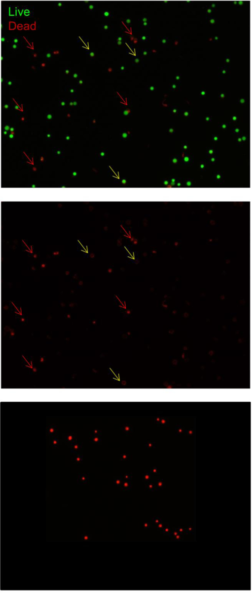
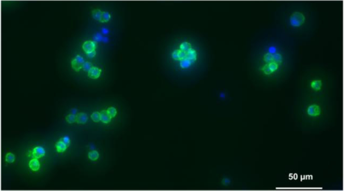
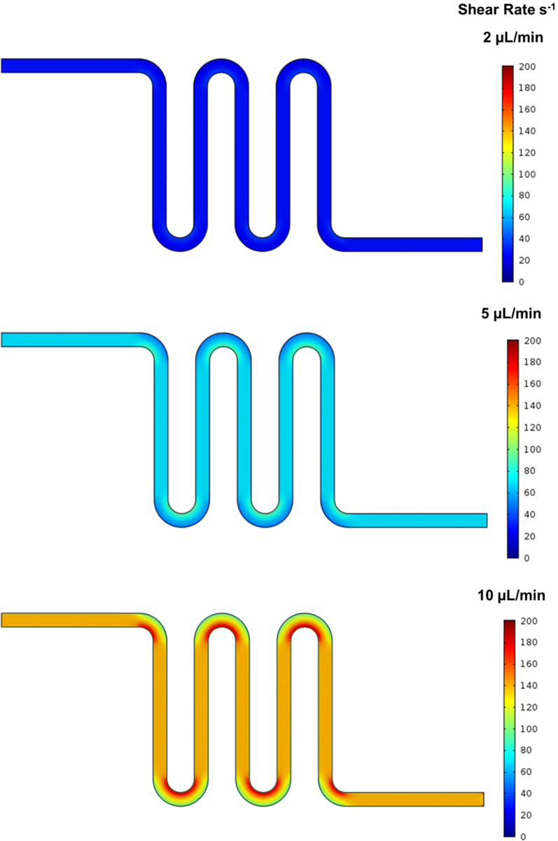
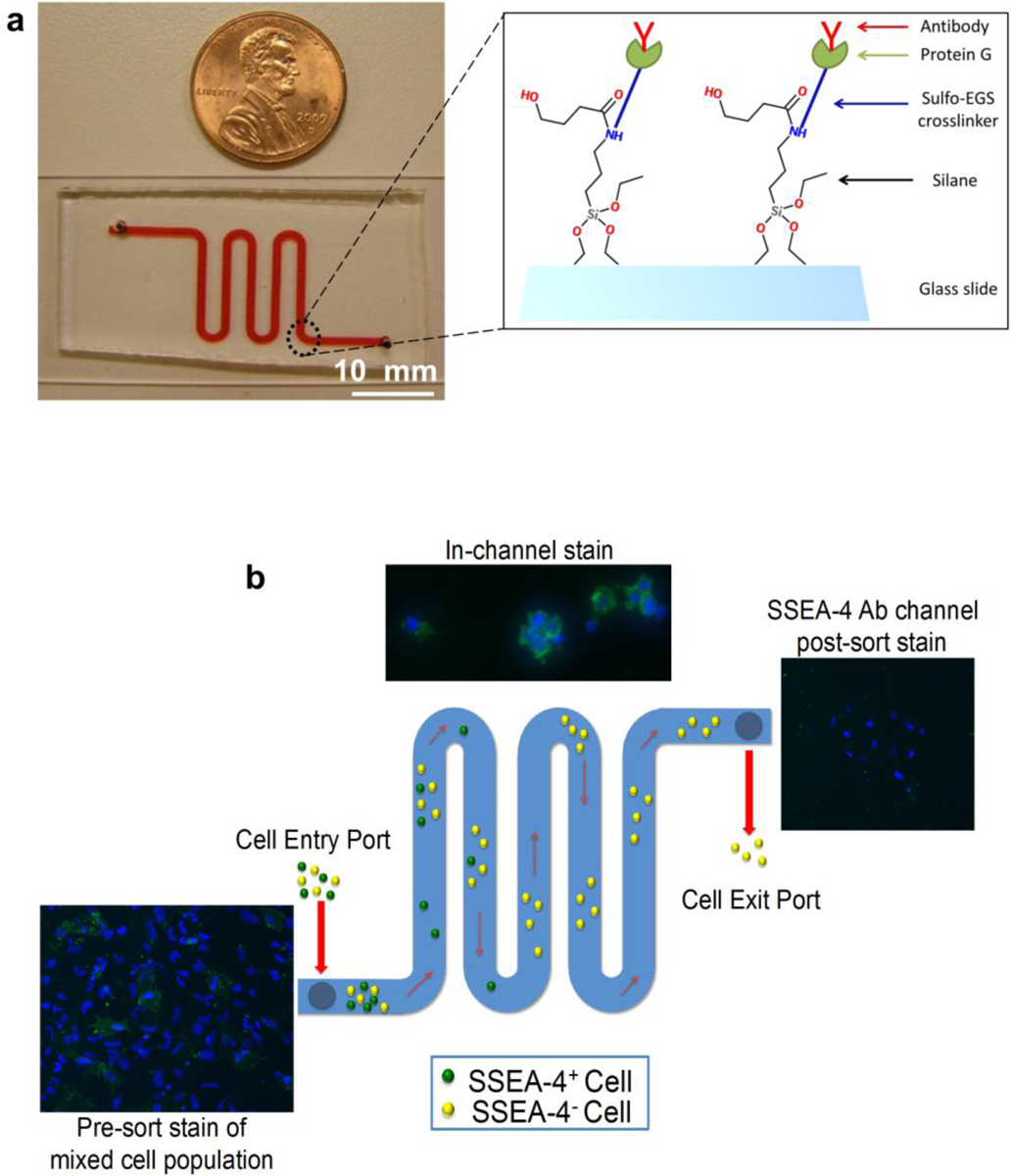
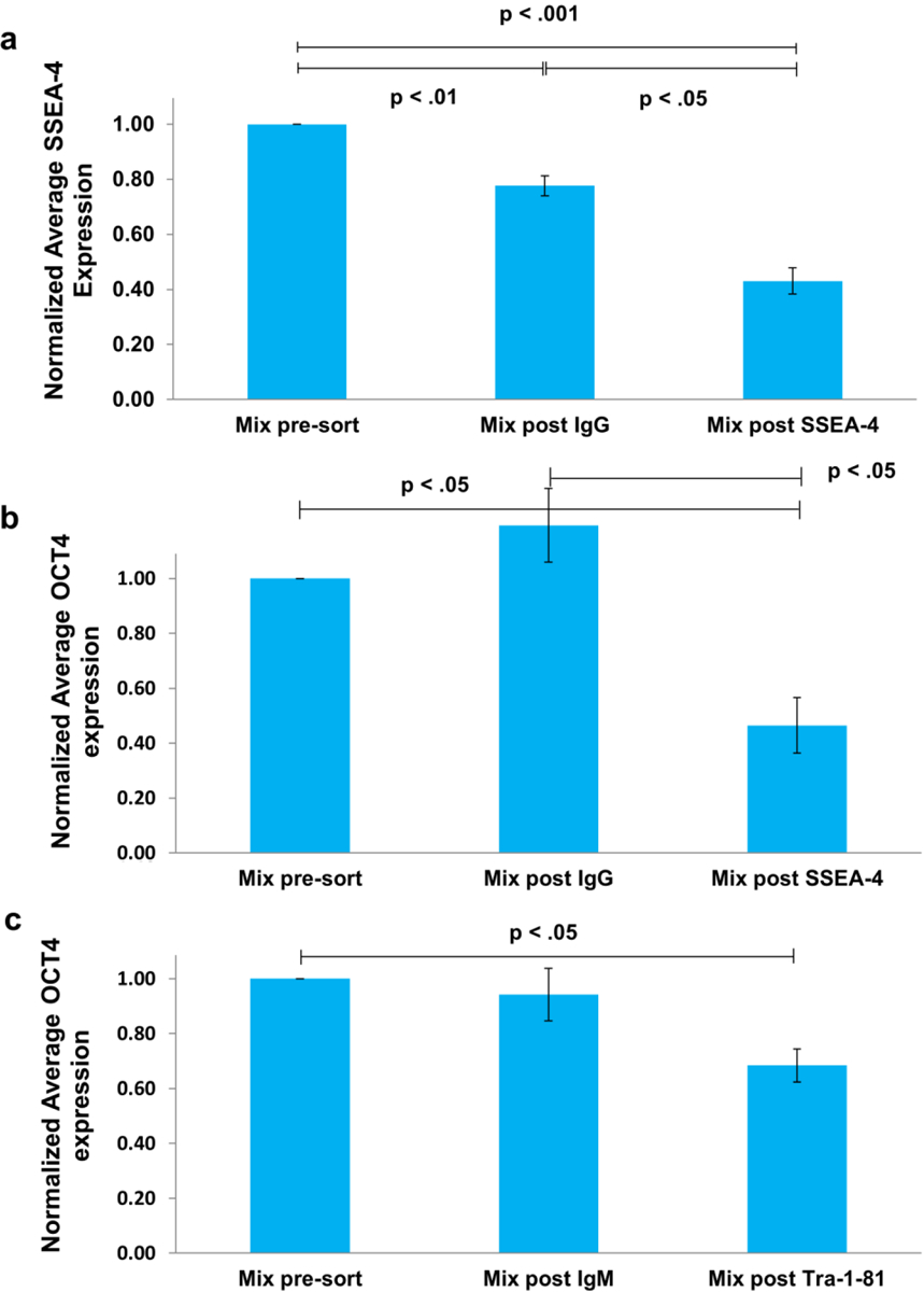
Conventional cell-sorting methods such as fluorescence-activated cell sorting (FACS) or magnetic-activated cell sorting (MACS) can suffer from certain shortcomings such as lengthy sample preparation time, cell modification through antibody labeling, and cell damage due to exposure to high shear forces or to attachment of superparamagnetic Microbeads. In light of these drawbacks, we have recently developed a label-free, microfluidic platform that can not only select cells with minimal sample preparation but also enable analysis of cells in situ. We demonstrate the utility of our platform by successfully isolating undifferentiated human embryonic stem cells (hESCs) from a heterogeneous population based on the undifferentiated stem-cell marker SSEA-4. Importantly, we show that, in contrast to MACS or FACS, cells isolated by our method have very high viability (~90%). Overall, our platform technology could likely be applied to other cell types beyond hESCs and to a variety of heterogeneous cell populations in order to select and analyze cells of interest.

Keywords: Human embryonic stem cells (hESCs); cell sorting; in situ analysis; label-free.


Figures

Fig.4
Fig.4
Fluorescent images of Live (green) / Dead (red) cells after SSEA-1+ FACS sorting. Cells labeled with PE (yellow arrows) show only faint, background fluorescence, along the cell membrane and are robustly green, i.e. live, whereas dead cells (red arrows) are bright and nuclear stain is clear, while the green, live signal is undetectable. The dead cell control image shows that ethidium homodimer 1 stained cells (red) do not have background levels of staining and are always bright and nuclearly-stained
Fig.5
Fig.5
hESCs bound inside an anti-SSEA-4-functionalized channel were stained for SSEA-4 (green) and Hoechst (blue). More than 90% of cells stained positive for SSEA-4
Fig.6
Fig.6
COMSOL-modeled shear rates in the microfluidic device at the running flow rate of 2 μL/min and the two washing flow rates, 5 μL/min and 10 μL/min
Fig.1
Fig.1
a) Image of the serpentine channel and inset of functionalization strategy (see Device functionalization). The (94 mm × 1 mm × 80 μm) (L × W × H) channel is cored at both ends for inlet and outlet ports. b) A schematic of the device method, using an anti-SSEA-4-functionalized-antibody channel as an example (although any antibody of choice can be used). A mixed population of cells is added at the inlet port. The outlet port is connected to a syringe pump via plastic tubing. As the cells traverse the device, SSEA-4+ cells become bound to the functionalized antibodies and SSEA-4 cells pass through the device and are collected in a syringe. At the end of the run, the device is washed thoroughly with fresh cell media, corresponding to the appropriate cell types (see Materials and Methods: Cell Culture).Control cells (i.e. those not injected into the channel) and uncaptured cells passed are plated, allowed to adhere to Matrigel-coated plates, fixed, and immunostained for either SSEA-4+ or OCT4+. Captured cells are stained and imaged in-channel. The percentage of SSEA-4+ or OCT4+ captured and not captured cells is then determined
Fig.2
Fig.2
Comparison of cell viability using MACS, FACS, and our microfluidics-based method. J1 mESCs cells were employed in all three methods and assayed for viability using a Live/Dead® assay. Average values from 3 independent experiments were normalized to results from cells that were maintained in media. Error bars corresponds to standard error. a) MACS: Assayed cells were collected under the following conditions: anti-IgM Microbeads (flow-through) and anti-SSEA-1 Microbeads (both flow-through and bound fractions). b) FACS: Assayed cells were those incubated with anti-IgM and those positively and negatively sorted for SSEA-1. c) Microfluidic isolation: Assayed cells were those that traversed anti-IgM- or anti-SSEA-1-functionalized antibody channels, as well as bound cells recovered from anti-SSEA-1-functionalized channels
Fig.3
Fig.3
a) Normalized average SSEA-4 expression for microchannel runs. A 4:1 ratio of hESCs to A549 were passed through either anti-IgG or anti-SSEA-4-functionalized channels. Control cells (those that do not run through any channel), and cells recovered after traversing either through anti-IgG channels or anti-SSEA-4 channels were plated and stained for the undifferentiated hESC marker, SSEA-4. Average values from 5 independent experiments were normalized to the fraction of SSEA-4 positive control cells. Error bars represent standard error. b) Normalized average OCT-4 expression for microchannel runs. A 4:1 ratio of hESCs to A549 cells were passed through either anti-IgG or anti-SSEA-4-functionalized channels. Control cells, cells recovered after traversing through anti-IgG channels, and cells recovered after passing through anti-SSEA-4 channels were plated and stained for the undifferentiated hESC antigen, OCT4. Average values from 4 independent experiments were normalized to the fraction of SSEA-4+ control cells. Error bars represent standard error. c) Normalized average OCT4 expression for microchannel runs. A 4:1 ratio of hESCs to A549s was passed through either anti-IgM or anti-Tra-1–81-functionalized channels. Control cells, cells recovered after traversing through anti-IgM channels, and cells recovered after passing through anti-Tra-1–81 channels were plated and stained for the undifferentiated hESC marker, OCT4. Average values from 3 independent experiments were normalized to the fraction of OCT4+ cells in the control cells. Error bars represent standard error

Similar articles

Cited by

References

    1. Abcam. (n.d.). Indirect flow cytometry (FACS) protocol. Retrieved from http://www.abcam.com/index.html?pageconfig=resource&rid=11381
    1. Abruzzese RV, & Fekete RA (2013). Chapter 17 Single Cell Gene Expression Analysis of Pluripotent Stem Cells. In Pluripotent Stem Cells (Vol. 997, pp. 217–224). doi:10.1007/978-1-62703-348-0 - DOI - PubMed
    1. Akerstroms B, & Bjork L (1989). Protein L : An Immunoglobulin Light Chain-binding Bacterial Protein, 264(33), 19740–19746. - PubMed
    1. Alix-Panabières C, & Pantel K (2014). Technologies for detection of circulating tumor cells: facts and vision. Lab on a Chip. doi:10.1039/c3lc50644d - DOI - PubMed
    1. Armstrong JK, Wenby RB, Meiselman HJ, & Fisher TC (2004). The hydrodynamic radii of macromolecules and their effect on red blood cell aggregation. Biophysical journal, 87(6), 4259–70. doi:10.1529/biophysj.104.047746 - DOI - PMC - PubMed