[Preprint]. 2021 Mar 2:2021.02.27.21252099.
doi: 10.1101/2021.02.27.21252099.

Persistent SARS-CoV-2 infection and increasing viral variants in children and young adults with impaired humoral immunity

Affiliations

Persistent SARS-CoV-2 infection and increasing viral variants in children and young adults with impaired humoral immunity

Thao T Truong et al. medRxiv. .

Update in

Abstract

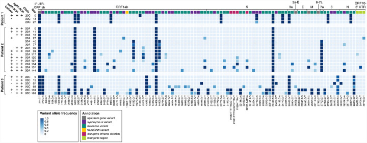
Background: There is increasing concern that persistent infection of SARS-CoV-2 within immunocompromised hosts could serve as a reservoir for mutation accumulation and subsequent emergence of novel strains with the potential to evade immune responses.

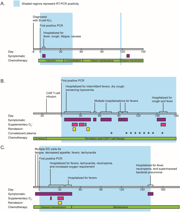
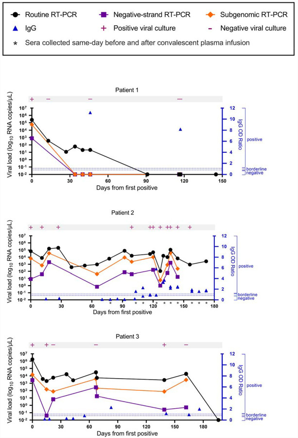
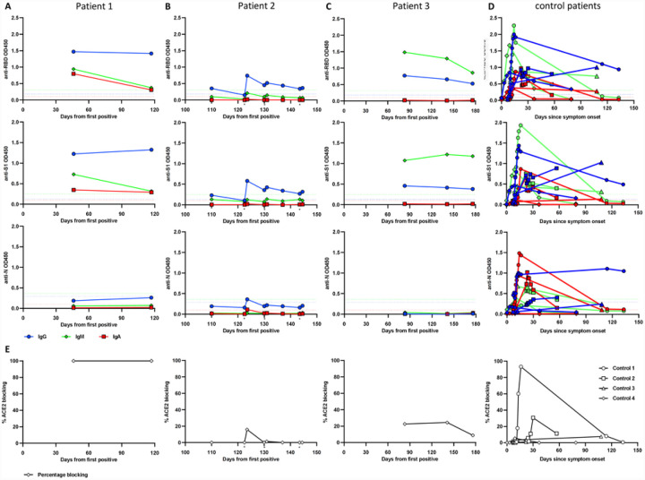
Methods: We describe three patients with acute lymphoblastic leukemia who were persistently positive for SARS-CoV-2 by real-time polymerase chain reaction. Viral viability from longitudinally-collected specimens was assessed. Whole-genome sequencing and serological studies were performed to measure viral evolution and evidence of immune escape.

Findings: We found compelling evidence of ongoing replication and infectivity for up to 162 days from initial positive by subgenomic RNA, single-stranded RNA, and viral culture analysis. Our results reveal a broad spectrum of infectivity, host immune responses, and accumulation of mutations, some with the potential for immune escape.

Interpretation: Our results highlight the need to reassess infection control precautions in the management and care of immunocompromised patients. Routine surveillance of mutations and evaluation of their potential impact on viral transmission and immune escape should be considered.

Funding: The work was partially funded by The Saban Research Institute at Children's Hospital Los Angeles intramural support for COVID-19 Directed Research (X.G. and J.D.B.), the Johns Hopkins Center of Excellence in Influenza Research and Surveillance HHSN272201400007C (A.P.), NIH/NIAID R01AI127877 (S.D.B.), NIH/NIAID R01AI130398 (S.D.B.), NIH 1U54CA260517 (S.D.B.), an endowment to S.D.B. from the Crown Family Foundation, an Early Postdoc.Mobility Fellowship Stipend to O.F.W. from the Swiss National Science Foundation (SNSF), and a Coulter COVID-19 Rapid Response Award to S.D.B. L.G. is a SHARE Research Fellow in Pediatric Hematology-Oncology.


Conflict of interest statement

Conflict of Interest S.D.B. has consulted for Regeneron, Sanofi, and Novartis on topics unrelated to this study. S.D.B., and K.R. have filed provisional patent applications related to serological tests for SARS-CoV-2 antibodies. All other authors have no competing interests.

Figures

Figure 1.
Figure 1.. Clinical timeline of symptoms, hospital admissions, and treatment.
Timelines for patient 1 (A), patient 2 (B), and patient 3 (C) are labelled by date from initial positive RT-PCR (day 0). Colored bars indicate time periods where patients were symptomatic, required supplementary oxygen, or received treatment (Remdesivir or convalescent plasma). The phases of chemotherapy are also shown.
Figure 2.
Figure 2.. Viral load by routine, negative-strand, and subgenomic RT-PCR.
Time course of viral load from nasopharyngeal or combined nares/oropharyngeal swabs collected from each patient. Viral culture results are indicated in pink. Corresponding serum anti-SARS-CoV-2 IgG values are plotted in blue. For patient 2, IgG was measured before and after administration of convalescent plasma at the indicated timepoints.
Figure 3.
Figure 3.. Anti-SARS-CoV-2 antibody responses in three immunocompromised pediatric and young adult patients.
Serum samples from two immunocompromised children (A, C) and one young adult (B) were collected up to 176 days after initial positive SARS-CoV-2 PCR test. Plasma samples from four non-immunocompromised COVID-19 patients were analyzed for comparison (D). Antibodies specific for SARS-CoV-2 Spike RBD (top row), S1 subunit (second row), and N protein (third row) were measured using ELISA at 1:100 serum/plasma dilution. RBD-ACE2 blocking activity was assessed in a competition ELISA and is shwon as percentage of blocking (E). The cutoff for seropositivity was defined as the mean absorbance + 3 SD of 94 pre-pandemic plasma samples in each assay (A, B, C, D). Color coding for isotypes: IgG (blue), IgM (green), IgA (red). Dotted lines depict the cutoff for seroconversion for the different isotypes in each assay. Individual donors were shown with different symbols (D). * indicates timepoints of convalescent plasma infusion in patient 2.
Figure 4.
Figure 4.. Accumulation of SARS-CoV-2 variants in three persistently positive pediatric and young adult patients with hematologic malignancies.
Each row represents viral culture, strand-specific RT-PCR (ssRT-PCR), subgenomic RT-PCR (sgRT-PCR) and sequencing from longitudinally derived specimens numbered by days from initial positive test (day 0). Boxes represent distinct variants, and shading reflects variant allele frequency (cutoff = 0.25). Corresponding genes are labeled in top row and colored to represent variant annotation (see legend). Specimens from two patients demonstrate progressive accumulation of multiple variants and variants of lower allele frequencies (Patient 2, difference of 7 major allele variants between days 0 and 144; Patient 3, difference of 13 major allele variants between days 0 and 162). In addition to point mutations, four different inframe deletions were observed to develop in the S gene in Patient 2 and Patient 3. Several variants (both major and minor alleles) reverted at later timepoints. UTR, untranslated region; S, spike; E, envelope; M, matrix; N, nucleocapsid

References

    1. Sethuraman N, Jeremiah SS, Ryo A. Interpreting Diagnostic Tests for SARS-CoV-2. JAMA 2020; 323: 2249. - PubMed
    1. CDC. COVID-19 and Your Health. Centers for Disease Control and Prevention. 2020; published online Feb 11. https://www.cdc.gov/coronavirus/2019-ncov/if-you-are-sick/quarantine.html (accessed Dec 27, 2020).
    1. Walsh KA, Spillane S, Comber L, et al. The duration of infectiousness of individuals infected with SARS-CoV-2. Journal of Infection 2020; 0. DOI:10.1016/j.jinf.2020.10.009. - DOI - PMC - PubMed
    1. Perera RAPM, Tso E, Tsang OTY, et al. SARS-CoV-2 Virus Culture and Subgenomic RNA for Respiratory Specimens from Patients with Mild Coronavirus Disease - Volume 26, Number 11—November 2020 - Emerging Infectious Diseases journal - CDC. DOI:10.3201/eid2611.203219. - DOI - PMC - PubMed
    1. Avanzato VA, Matson MJ, Seifert SN, et al. Case Study: Prolonged infectious SARS-CoV-2 shedding from an asymptomatic immunocompromised cancer patient. Cell 2020; published online Nov 4. DOI:10.1016/j.cell.2020.10.049. - DOI - PMC - PubMed

Publication types

LinkOut - more resources