

Imaging diagnosis of classical and new pneumoconiosis: predominant reticular HRCT pattern
- PMID: 33689008
- PMCID: PMC7947097
- DOI: 10.1186/s13244-021-00966-y
Imaging diagnosis of classical and new pneumoconiosis: predominant reticular HRCT pattern
Abstract
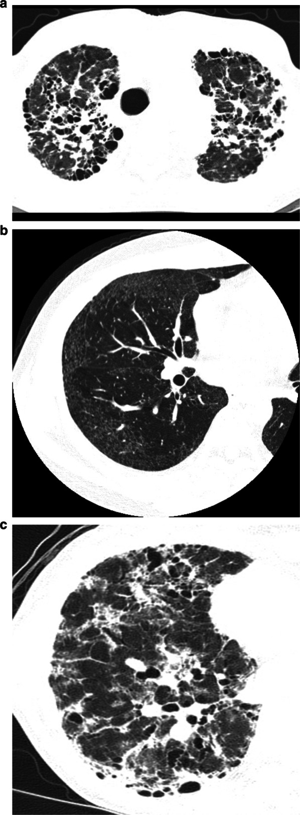
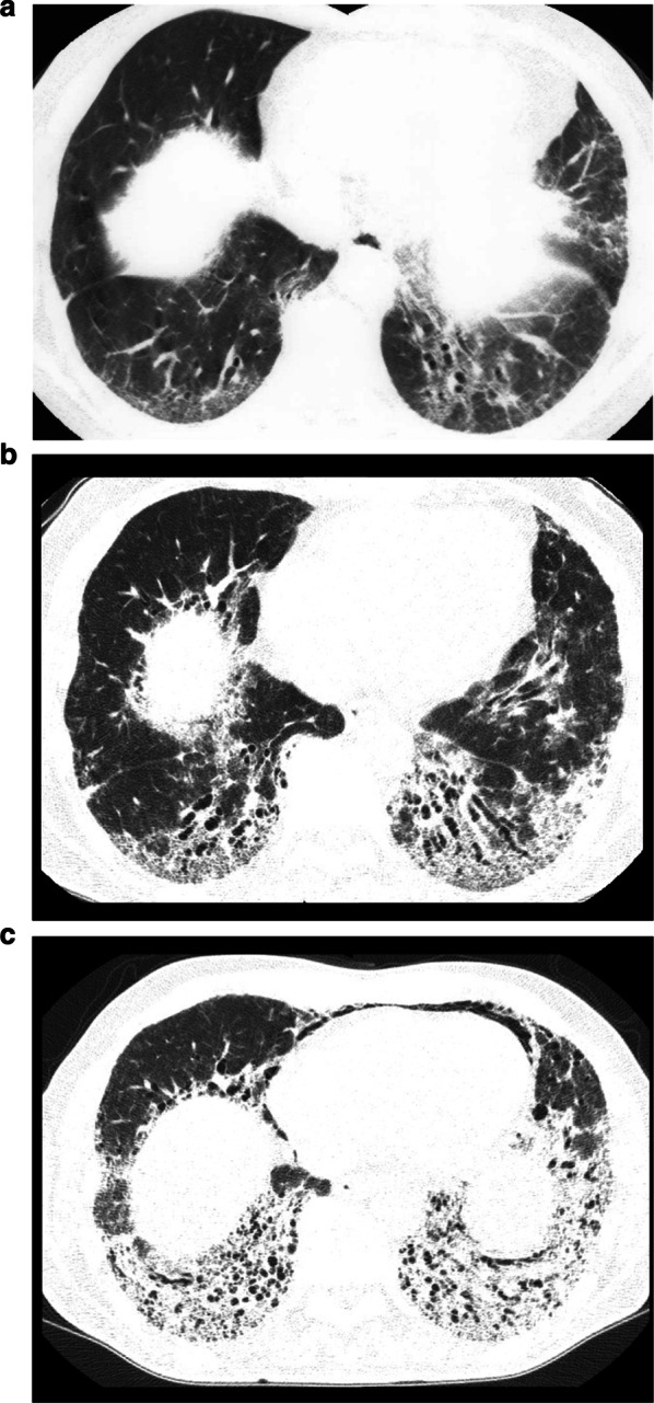
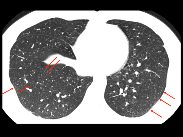
Our understanding of the manifestations of pneumoconioses is evolving in recent years. Associations between novel exposures and diffuse interstitial lung disease have been newly recognized. In advanced asbestosis, two types of fibrosis are seen, probably related to dose of exposure, existence of pleural fibrosis, and the host factor status of the individual. In pneumoconiosis of predominant reticular type, nodular opacities are often seen in the early phase. The nodular pattern is centrilobular, although some in metal lung show perilymphatic distribution, mimicking sarcoidosis. High-resolution computed tomography enables a more comprehensive correlation between the pathologic findings and clinically relevant imaging findings. The clinician must understand the spectrum of characteristic imaging features related to both known dust exposures and to historically recent new dust exposures.
Keywords: Asbestosis; HRCT; Metal lung disease; Pneumoconiosis.
Conflict of interest statement
The author declares that he/she have no competing interests.
Figures
Similar articles
-
Imaging diagnosis of pneumoconiosis with predominant nodular pattern: HRCT and pathologic findings.Clin Imaging. 2023 May;97:28-33. doi: 10.1016/j.clinimag.2023.02.010. Epub 2023 Feb 18. Clin Imaging. 2023. PMID: 36878176 Review.
-
Pneumoconiosis: comparison of imaging and pathologic findings.Radiographics. 2006 Jan-Feb;26(1):59-77. doi: 10.1148/rg.261055070. Radiographics. 2006. PMID: 16418244 Review.
-
Thoracic Sarcoidosis: Imaging with High Resolution Computed Tomography.J Clin Diagn Res. 2017 Feb;11(2):TC15-TC18. doi: 10.7860/JCDR/2017/24165.9459. Epub 2017 Feb 1. J Clin Diagn Res. 2017. PMID: 28384959 Free PMC article.
-
Differentiation of etiology of nodular changes in high resolution computed tomography (HRCT) in interstitial lung diseases.Ann Univ Mariae Curie Sklodowska Med. 2003;58(2):370-7. Ann Univ Mariae Curie Sklodowska Med. 2003. PMID: 15323221
-
[Pathologic-HRCT correlation of pneumoconiosis--a study on inflation-fixed lungs].Nihon Igaku Hoshasen Gakkai Zasshi. 1992 Jan 25;52(1):35-51. Nihon Igaku Hoshasen Gakkai Zasshi. 1992. PMID: 1549447 Japanese.
Cited by
-
Impact of coal mine dust exposure and cigarette smoking on lung disease in Appalachian coalminers.Respir Res. 2025 May 14;26(1):184. doi: 10.1186/s12931-025-03260-3. Respir Res. 2025. PMID: 40369555 Free PMC article.
-
Early Identification, Accurate Diagnosis, and Treatment of Silicosis.Can Respir J. 2022 Apr 25;2022:3769134. doi: 10.1155/2022/3769134. eCollection 2022. Can Respir J. 2022. PMID: 35509892 Free PMC article. Review.
-
[Occupational interstitial lung diseases].Radiologie (Heidelb). 2024 Aug;64(8):636-642. doi: 10.1007/s00117-024-01342-9. Epub 2024 Jul 16. Radiologie (Heidelb). 2024. PMID: 39012478 Free PMC article. Review. German.
-
Survival analysis of patients with pneumoconiosis followed in occupational medicine clinics: 10 years experience.BMC Pulm Med. 2025 May 16;25(1):236. doi: 10.1186/s12890-025-03676-z. BMC Pulm Med. 2025. PMID: 40380112 Free PMC article.
-
Brazilian Thoracic Society recommendations for the diagnosis and monitoring of asbestos-exposed individuals.J Bras Pneumol. 2024 Aug 19;50(3):e20240156. doi: 10.36416/1806-3756/e20240156. eCollection 2024. J Bras Pneumol. 2024. PMID: 39166593 Free PMC article.
References
Publication types
LinkOut - more resources
Full Text Sources
Other Literature Sources
